

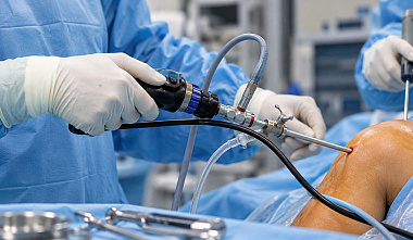
When a joint is injured, a doctor may need to see the condition of the tissues from the inside rather than assess them only through indirect signs. Arthroscopic examination is used for this purpose. An optical endoscope is inserted into the joint cavity through a small surgical access, and the image is transmitted to a monitor. This allows the specialist to examine intra-articular structures and, if necessary, immediately proceed with a therapeutic procedure.
What Is an Arthroscope and What Is It Used For?
An arthroscope is a medical endoscope used for diagnosing and performing surgery on joints. It is a thin, rigid optical system connected to a camera, light source, and other components of an endoscopic system. Depending on the model, the equipment may differ in diameter, length, viewing angle, and design. Its primary purpose is to provide visual control of the joint cavity. Unlike external examination or imaging methods, arthroscopy provides the doctor with a direct view of the internal tissues.
The method is used for:
-
joint injuries;
-
chronic pain and limited mobility;
-
cartilage damage;
-
tears and other meniscal injuries;
-
ligament injuries;
-
inflammatory changes;
-
other diseases of the musculoskeletal system.
When indicated, the diagnostic procedure can be combined with surgical treatment. Through separate access points, the doctor inserts specialized instruments and performs the necessary procedures:
-
removes damaged tissue fragments;
-
treats the cartilage surface;
-
removes loose bodies from the joint cavity;
-
repairs damaged structures;
-
performs suturing of certain tissues.
The specific scope of arthroscopy is determined by the nature of the disease, the location of the injury, and the condition of the joint.
Types of Arthroscopes
When selecting an arthroscope, not only its optical characteristics but also the requirements of the planned examination or surgery are taken into account. The diameter, viewing angle, working length, and image transmission method determine the convenience of use for the surgeon and the capabilities of the equipment. ElePS offers arthroscopes for different clinical applications, allowing a model to be selected according to the specific area of use and the requirements of the endoscopic system.
The main types of equipment can be classified according to several characteristics:
-
by viewing angle — 0°, 30°, 70°, and other configurations;
-
by diameter — standard models of approximately 4 mm and thinner arthroscopes with a diameter of 2.7 mm;
-
by purpose — diagnostic and operative models designed for use as part of a surgical system;
-
by image transmission method — optical arthroscopes with an eyepiece and models connected to a video system.
The viewing angle is selected according to the anatomical area and the intended procedures. 0° optics provide a direct view along the insertion axis, 30° optics expand the field of view forward and to the sides, while 70° optics help examine areas located to the side of the insertion direction. Arthroscopes with a diameter of 2.7 mm are suitable when a smaller access point is required, while 4 mm models provide the conventional visualization format used in standard arthroscopic procedures.
ElePS offers arthroscopes with diameters of 2.7 and 4 mm and viewing angles of 0°, 30°, and 70°. The catalog allows users to select optics with the required parameters for diagnostic or surgical arthroscopy and choose equipment compatible with the endoscopic video system in use.
Flexible and Rigid Arthroscopes
By design, endoscopes are divided into rigid and flexible types. Rigid models are used most commonly in arthroscopy. Their optical system is enclosed in a metal housing that maintains a fixed geometry during the procedure. This design provides stable positioning of the optics and accurate image transmission.
Flexible endoscopes have a movable working section and a controllable distal tip. They are used considerably less frequently in joint surgery because most standard arthroscopic procedures are performed using rigid optics.
Arthroscope Design and Components
The design of an arthroscope determines the capabilities of the equipment during diagnostic examinations and surgical procedures. A rigid endoscope consists of an optical system enclosed in a metal housing. It is based on an objective lens and a lens system that produces an image inside the joint. The housing has connectors for the light guide and camera, while a specialized sheath is used to safely introduce the optics.
Depending on the requirements of the procedure, an arthroscopic system may include:
-
optics with the selected diameter and viewing angle;
-
sheaths of different diameters;
-
trocars and stylets;
-
an endoscopic camera and video system;
-
a cold light source;
-
a light guide cable;
-
an irrigation pump;
-
surgical instruments.
The optics transmit the image from the joint cavity, the camera converts it into a video signal, and the video system displays the image on a medical monitor. Modern systems can provide high-resolution visualization as well as photo and video recording of the procedure.
The equipment configuration is selected according to the clinic's area of specialization and the types of procedures performed. Probes, forceps, scissors, shavers, electrodes, and suture systems are used for diagnostic procedures and surgeries involving the menisci, cartilage, ligaments, and other structures.
ElePS, a Russian manufacturer of endoscopic equipment, supplies components for arthroscopic systems, including optics and related equipment. When selecting equipment, factors such as the arthroscope diameter and viewing angle, connection format, and compatibility between components can be taken into account. This is particularly important when upgrading an existing endoscopic system, as the equipment can be selected to match the existing configuration without replacing the entire system.
How an Arthroscope Works
Before the procedure, the surgeon creates a small access point to the joint cavity. A sheath containing the optical system is inserted through this access point. Light is delivered to the examination area through a light guide. The image from the objective lens is transmitted to the camera and then displayed on a monitor. The surgeon controls the position of the endoscope and systematically examines the accessible intra-articular structures. To improve visualization, sterile fluid is introduced into the joint. It expands the space and simultaneously helps remove blood and small particles from the working area.
If surgery is required, specialized instruments are inserted through additional puncture sites. The surgeon controls their position using the image displayed on the monitor. This makes it possible to perform diagnosis and treatment during a single arthroscopic procedure.
Which Joints Can Be Examined Using an Arthroscope?
The method is used in various areas of orthopedics and traumatology. Arthroscopic procedures are most commonly performed on:
-
the knee joint, for meniscal, ligament, and cartilage injuries and loose intra-articular bodies;
-
the shoulder joint, for injuries of the rotator cuff, glenoid labrum, and other structures;
-
the hip joint, for the diagnosis and treatment of cartilage and labral injuries;
-
the ankle joint, for cartilage injuries, inflammatory changes, and certain consequences of trauma;
-
the elbow joint, for certain cartilage injuries, loose bodies, and other intra-articular pathologies;
-
the joints of the hand and foot, when appropriate indications are present and suitable equipment is available.
The diameter of the optics and the set of instruments are selected according to the anatomical area. Therefore, a system designed for a large knee joint may differ from the equipment used for a small joint of the hand.
What Can Be Seen Using an Arthroscope?
During the examination, the doctor obtains direct visual access to structures inside the joint. The field of view may include:
-
articular cartilage;
-
menisci;
-
ligaments;
-
synovial membrane;
-
joint capsule;
-
certain areas of tendons;
-
loose fragments of bone or cartilage;
-
areas of inflammation and tissue damage.
High image quality is particularly important when working with small anatomical structures. The ability to magnify and record video helps document the course of the procedure and the treatment results.
How Arthroscopy Is Performed
The procedure is performed in an operating room and includes several successive stages:
-
Preparation. The doctor assesses the patient's condition, confirms the diagnosis, and determines the scope of the surgical procedure.
-
Anesthesia. An appropriate type of anesthesia is selected based on the procedure and the patient's condition.
-
Creating access. A small puncture or incision is made in the area of the joint.
-
Optics insertion. A sheath containing the optical system is inserted into the joint.
-
Irrigation. Sterile solution is introduced into the joint cavity to expand the space and improve visualization.
-
Examination. The surgeon examines the intra-articular structures using the image displayed on the monitor.
-
Treatment. Specialized instruments are inserted through additional access points and the necessary procedures are performed.
-
Completion. The instruments and optics are removed, after which the surgeon cleans and closes the surgical access points.
A complete arthroscopic system requires optics, a camera, a light source, a monitor, an irrigation system, and surgical instruments. ElePS offers equipment for endoscopic surgery, including arthroscopes and components for arthroscopic systems. When configuring the system, the specifications of the particular model, component compatibility, and the requirements of the medical facility are taken into account.


