

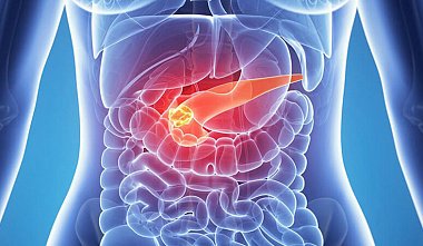
Destructive pancreatitis: diagnostic and treatment algorithm
Introduction: Patients with acute pancreatitis account for 5-10% of all surgical patients. In 15-20% of cases, acute pancreatitis is destructive. In 40-70% of patients with pancreatic necrosis, infection of the necrotic destruction foci occurs. Infectious complications account for 80% of deaths in patients with destructive pancreatitis. The purpose of this document is to develop a unified classification and a standardized algorithm for the diagnosis, examination, and treatment of destructive pancreatitis. However, this project cannot cover all aspects of this significant problem. The document is based on the experience of surgical clinics in the Russian Federation and data from international evidence-based studies on destructive pancreatitis. Clinical and morphological classification of acute pancreatitis. The proposed classification corresponds to the current level of comprehensive clinical, laboratory, and topical instrumental diagnostics of acute pancreatitis and its various complications. It is based on the understanding of the staged transformation of necrotic destruction zones and the development of complications depending on the prevalence and nature of the lesion of the pancreas (PG) and retroperitoneal tissue under the influence of endogenous and exogenous infection factors. Forms of acute pancreatitis - Edematous (interstitial) pancreatitis. - Necrotic pancreatitis (pancreatic necrosis) sterile: by the nature of the necrotic lesion: - fatty, - hemorrhagic, - mixed; by the prevalence of the lesion: - small focal, - large focal; by localization: - with damage to the head, - body, - tail, - all parts of the pancreas. - Necrotic pancreatitis (pancreatic necrosis) infected. Complications of acute pancreatitis - Parapancreatic infiltrate. - Pancreatogenic abscess. - Peritonitis: enzymatic (abacterial), bacterial. — Septic phlegmon of the retroperitoneal tissue: - parapancreatic, - paracolic, - perinephric, - pelvic. — Erosive bleeding. — Mechanical jaundice. — Pseudocyst: - sterile, - infected. — Internal and external fistulas of the gastrointestinal tract. The main clinical and morphological form of destructive pancreatitis in the early stages of the disease is sterile pancreatic necrosis. Infected forms of pancreatic necrosis are detected on average in 25% of patients in the 1st week, in 30% - in the 2nd week, and in 70% - in the 3rd week of the disease. Pancreatogenic abscesses, as a rule, develop in periods of more than 3 months of the disease. Diagnosis of acute pancreatitis To improve the quality of diagnosis and determine the prognosis for acute pancreatitis, first of all, it is necessary to thoroughly evaluate the clinical picture of the disease, determine the activity of pancreatic enzymes in the blood (amylase, lipase), urine (amylase), peritoneal exudate, the dynamics of homeostatic parameters (blood leukocytes, leukocyte intoxication index, hematocrit, glucose, bilirubin, urea, creatinine, total protein, albumin, ALT, AST, LDH, Na, K, Ca, Cl, PA07 in the blood), as well as a comprehensive instrumental examination, including ultrasound data (ultrasound) of the retroperitoneal organs and abdominal cavity, laparoscopy, computed tomography (CT). The diagnosis of acute pancreatitis should be verified within the first 2 days of hospitalization of the patient in a surgical hospital. Chest and abdominal radiography allows for differential diagnosis with perforation of a hollow organ, mechanical intestinal obstruction, and the presence of acute lung injury (atelectasis, pneumonia, ARDS, concomitant pleurisy). Ultrasound is a mandatory screening method for assessing the condition of the pancreas, biliary system, abdominal and pleural cavities in acute pancreatitis. Ultrasound allows for the diagnosis of acute pancreatitis in 40-85% of cases, but does not always reliably verify the form of acute pancreatitis or characterize the condition of the retroperitoneal fat. Measuring hemodynamic parameters in visceral vessels, pancreatic density, and retroperitoneal fat increases the specificity, sensitivity, and accuracy of ultrasound in pancreatic necrosis. Laparoscopy is a mandatory diagnostic and therapeutic method. However, this method does not always allow for direct examination of the pancreas or assessment of the extent and nature of damage to the pancreas, peritoneum, and retroperitoneal fat. Videolaparoscopy allows for decompressive gallbladder surgery, necrosequestrectomy through an omentopancreatobursostomy, and dynamic laparoscopy and abdominal debridement in cases of pancreatogenic peritonitis. Indications for therapeutic and diagnostic laparoscopy: — Differential diagnosis of peritonitis of various etiologies. — Clinically diagnosed papreonecrosis for verification of its form and drainage (lavage) of the abdominal cavity in cases of enzymatic peritonitis. — Laparoscopic cholecystostomy for biliary hypertension. CT is the "gold standard" and the most sensitive imaging technique for acute pancreatitis and its complications, providing comprehensive information on the condition of the pancreas and retroperitoneal space, including the involvement of the biliary tract, underlying vascular structures, and gastrointestinal tract. Indications for CT in acute pancreatitis: — Verification of the clinical form of acute pancreatitis when there is insufficient information from clinical, laboratory, and instrumental (ultrasound, laparoscopy) data. — Assessment of the prevalence and nature of the lesion of the pancreas and various parts of the retroperitoneal tissue in papillopecrosis within 3-10 days from the moment of hospitalization (the timing of the study is determined by the optimal time for demarcation development in the pancreas). — Worsening of the patient's condition due to suspected development of complicated forms. — The presence of a palpable infiltrate in combination with signs of a systemic inflammatory reaction and intoxication. — For planning and performing transcutaneous diagnostic and therapeutic punctures and/or drainage of fluid formations in the retroperitoneal localization. — To determine a rational surgical approach and planning the extent of surgical intervention. CT data form the basis for an objective assessment of the severity of acute pancreatitis. Contrast-enhanced CT (pancreatoangioscanning) allows for highly accurate diagnosis of pancreatic necrosis, assessment of its extent and localization, and detection of various angiogenic complications (erosion of pancreatic and parapancreatic vessels, pseudoaneurysm formation, occlusion of portal vein branches). Five severity grades of the disease should be distinguished based on contrast-enhanced CT data, which can be summarized in the form of a scale for integral assessment of the extent and nature of damage in acute pancreatitis: A) Normal pancreas (0 points). B) Local or diffuse enlargement of the pancreas in combination with hypodense inclusions in its tissue with fuzzy edges, dilation of the pancreatic duct (1 point). C) Changes in pancreatic tissue similar to stage B, to which inflammatory changes are added in the parapancreatic tissue (2 points). D) Changes C + isolated fluid formations outside the pancreas (3 points). E) Changes D + two or more fluid formations outside the pancreas or the presence of an abscess - gas formation (4 points). CT and ultrasound allow differentiation of fluid formations from soft tissue inflammatory-necrotic masses, but do not provide a diagnosis of sterile and infected destruction. Therefore, a method for early and accurate differential diagnosis of sterile pancreatic necrosis and its septic complications is percutaneous puncture under ultrasound or CT guidance with immediate Gram staining of the biosubstrate smear, followed by bacteriological examination to determine the type of microorganism and its sensitivity to antibiotics. Endoscopic visualization of the ampulla of Vater and retrograde cholangiopancreatography (ERCP), and, if necessary, endoscopic papillotomy are indicated in biliary pancreatitis with mechanical jaundice and/or cholangitis, taking into account the detection of a dilated common bile duct in diameter, according to ultrasound data, and the ineffectiveness of complex conservative therapy within 48 hours. Severity assessment An objective assessment of the severity of acute pancreatitis, including three main stages, should be performed in all patients within the first 2 days of hospitalization. Primary (initial) assessment of the severity of acute pancreatitis involves clinical differentiation into the interstitial form of the disease and pancreatic necrosis based on the severity of local symptoms from the abdominal organs and their correspondence to systemic organ disorders (cardiac, respiratory, renal, hepatic, intestinal, cerebral, metabolic failure). The accuracy and prognostic value in this case are only 50%. At the second stage, the assessment of the severity of the disease is based on the analysis of several clinical and laboratory scales for the integrated assessment of the parameters of the physiological state of the patient with acute pancreatitis, which can increase the accuracy of predicting the outcome of the disease and the development of postnecrotic complications to 70-80%. The most common systems for the integrated assessment of the severity of the patient's condition and the prognosis of acute pancreatitis are the Ranson (1974), Glasgow (1984), and APACHE II (1984) scales. With Ranson/Glasgow scale values ????more than 4 points, and APACHE II more than 9 points, the development of destructive pancreatitis is predominantly complicated. Dynamic daily assessment of the severity of the patient's condition using the APACHE II scale forms the basis for objectifying indications for surgery and a differentiated approach to choosing a comprehensive treatment strategy for pancreatic scoliosis. The use of integrated scales allows for an objective assessment of the results of various treatment methods, which is particularly important for comparative studies in the Russian Federation. Currently, to accurately predict the course of acute pancreatitis and its complications, it is advisable to determine transaminase activity, C-reactive protein levels, and, if possible, procalcitonin in the blood of patients with pancreatitis over the course of the disease. In the third stage, disease severity assessment is based on determining the extent and nature of pancreatic, retroperitoneal, and abdominal lesions during the diagnosis of pancreatic necrosis based on ultrasound, laparoscopy, and CT scan results. Treatment strategies for acute pancreatitis. The tactics and methods of comprehensive treatment for destructive pancreatitis are determined by the phase of the pathological process and the severity of the patient's condition. Comprehensive treatment of patients with destructive pancreatitis should be performed in an intensive care unit. Main approaches and methods of comprehensive treatment for destructive pancreatitis. Intensive care is aimed at restoring hemodynamics, rehydration, correcting acid-base balance disorders, and maintaining optimal oxygen delivery. Blockade of pancreatic secretory function and mediatosis. For this purpose, it is primarily advisable to use octreotide at a dose of 100 mcg 3 times daily for 5-7 days or antimetabolites (5-fluorouracil) at a dose of 10 mg/kg once, or less often twice. The lack of evidence on the efficacy of protease inhibitors in pancreatic necrosis does not allow us to recommend their further clinical use at this time. Extracorporeal detoxification. Antibacterial prophylaxis and therapy. The choice of antibacterial drugs for pancreatic necrosis is based on microbiological studies. The spectrum of antibiotic activity should cover gram-negative and gram-positive aerobic and anaerobic microorganisms that cause pancreatogenic infections. This ensures a rational empirical regimen of antimicrobial prophylaxis and therapy for pancreatic necrosis. The second determinant of antibiotic efficacy is their ability to selectively penetrate pancreatic tissue through the hematopancreatic barrier. Three groups of antibacterial drugs can be distinguished depending on their different penetrating abilities into pancreatic tissue. Group I. Aminoglycosides, aminopenicillins, and first-generation cephalosporins, the concentration of which after intravenous administration does not reach the minimum inhibitory concentration (MIC) in pancreatic tissue for most bacteria. Group II is represented by antibacterial drugs, the tissue concentration of which after intravenous administration exceeds the MIC for many microorganisms encountered in pancreatic infection - protected broad-spectrum penicillins: piperacillin/tazobactam and ticarcillin/clavulanate; third-generation cephalosporins: cefoperazone and cefotaxime; fourth-generation cephalosporins (cefepime). Group III includes fluoroquinolones (ciprofloxacin and especially pefloxacin) and carbapenems (imipenem/cilastatin, meropenem), which produce concentrations in pancreatic tissues significantly exceeding the MIC for most pathogens associated with pancreatic necrosis. Metronidazole also achieves bactericidal concentrations in pancreatic tissues for anaerobic bacteria and can therefore be used as a component of combination antibacterial therapy (third- or fourth-generation cephalosporin + metronidazole). Antibiotic use in acute pancreatitis: Antibacterial prophylaxis is not indicated for edematous pancreatitis. A diagnosis of pancreatic necrosis is an absolute indication for the use of antibacterial drugs (groups II and III), which produce effective bactericidal concentrations in the affected area. In many cases, it is difficult to immediately differentiate between the prophylactic and therapeutic purposes of antibiotic administration for pancreatic necrosis, given the high risk of infection of the necrotic pancreas and the difficulty of determining it using clinical, laboratory, and instrumental methods in real time. The development of fatal sepsis in pancreatic necrosis requires the immediate administration of antibacterial drugs with maximum efficacy and minimal side effects. Efficacy should take precedence over cost. The drugs of choice for both prophylactic and therapeutic use in pancreatic necrosis are: - carbapenems, - fluoroquinolones (especially pefloxacin) + metronidazole, - third- and fourth-generation cephalosporins + metronidazole, - protected penicillins (piperacillin/tazobactam, ticarcillin/clavulanate). The duration of antibacterial therapy for pancreatic necrosis is determined by the time it takes for the symptoms of the systemic inflammatory reaction to completely resolve. Considering the important role of intestinal bacterial translocation in the pathogenesis of infectious complications of pancreatic necrosis, it is advisable to include a regimen of selective intestinal decontamination, in particular, the oral administration of aminoglycosides (gentamicin/tobramycin 320 mg/day + polymyxin M 400 mg/day) or fluoroquinolones (pefloxacin 800 mg/day). Pancreatic necrosis is a risk factor for the development of fungal superinfection, which determines the appropriateness of including antifungal agents (fluconazole) in the treatment program for patients with pancreatic necrosis. Given the dynamics of the pathological process in pancreatic necrosis from sterile to infected and the often multi-stage nature of surgical interventions, for effective antibacterial therapy, it is necessary to provide for a change in its regimen based on clinical and laboratory signs, rather than temporary ones. Nutritional support is indicated for patients with severe acute pancreatitis (Ranson score >2, APACHE II score >9), and with a confirmed clinical diagnosis of pancreatic necrosis. If edematous pancreatitis is diagnosed and there is a positive response to comprehensive treatment within 48-72 hours, a transition to a natural diet according to diets 1 and 5 is indicated after 5-7 days. In cases of pancreatic necrosis, oral nutrition should be discontinued and artificial nutrition should be used. The effectiveness of long-term (2-3 weeks) total parenteral nutrition in pancreatic necrosis is questionable. This is explained by the following adverse effects: enterocyte atrophy, increased contamination and enterogenous translocation of bacteria and microbial toxins, the risk of developing angiogenic infection and cholestasis, immunosuppression, and the high cost of this nutritional method. В этой связи более целесообразным и эффективным при панкреонекрозе считают переход на энтеральное искусственное питание в ранние сроки заболевания. Нутритивную поддержку осуществляют через назоеюнальный зонд установленный дистальнее связки Трейтца эндоскопическим способом или во время операции. Питательные смеси - стандартные: берламин, нутризон, нутрилан и др. Режим энтерального питания -стартовый по темпу и объему, с постепенным переходом от глюкозо-солевых растворов и химусоподобных смесей к олигомерным питательным смесям в возрастающих концентрациях 5-10-20%. В случае развития непереносимости энтерального питания (увеличение уровней амилаз- и липаземии, стойкий парез кишечника, диарея, аспирация) у больных панкреонекрозом показан переход на полное парентеральное питание с возвратом к энтеральному питанию по мере восстановления функции желудочно-кишечного тракта. Хирургическое лечение панкреонекроза В отношении принципов дифференцированного хирургического лечения панкреонекроза и его гнойно-септических осложнений имеются принципиальные различия. Они касаются оптимальных сроков и режимов оперативного вмешательства, доступов, видов операций на ПЖ, желчевыводящей системе, методов дренирующих операций забрюшинного пространства и брюшной полости. Показанием к операции при панкреонекрозе является. Инфицированный панкреонекроз и/или панкреатогенный абсцесс, септическая флегмона забрюшинной клетчатки, гнойный перитонит независимо от степени полиорганных нарушений. Факт инфицирования некротических тканей является важным, но не единственным показанием к операции, особенно в ранние сроки заболевания. Стойкая или прогрессирующая полиорганная недостаточность, несмотря на комплексную интенсивную консервативную терапию в течение 1-3 сут, что свидетельствует об обширном некрозе ПЖ и забрюшинной клетчатки или высоком риске развития панкреатогенной инфекции. Для объективизации показаний к операции необходимо использовать интегральные шкалы оценки тяжести состояния больного (Ranson/Glasgow, APACHE II), распространенности и характера поражения ПЖ и забрюшинной клетчатки (шкала КТ). Оперативное лечение показано больным, у которых, по данным КТ с контрастным усилением, распространенность некроза превышает 50% паренхимы ПЖ и/или диагностировано обширное распространение некроза на забрюшинное пространство, что соответствует высокому риску инфицирования и фатальных системных осложнений. Панкреатогенный (ферментативный, абактериальный) перитонит является показанием к лапароскопиче-ской санации и дренированию брюшной полости. Хирургические доступы: срединная или 2-подреберная лапаротомия. Методы хирургического лечения варьируют, что определяется динамикой патоморфологического процесса в ПЖ, забрюшинной клетчатке и брюшной полости. Техническое решение этапа некрсеквестрэктомии однотипно и предполагает ее выполнение в максимально возможном объеме с сохранением естественных отграничивающих структур - брюшины мезентериального синуса и брыжейки ободочной кишки. Особое значение необходимо придавать мобилизации соответствующих сегментов ободочной кишки по латеральному контуру и выбору метода дренирующих операций в зависимости от распространенности поражения ПЖ и различных отделов забрюшинного пространства. Избранный уже на первой операции метод дренирующих операций существенно определяет выбор режима оперативной тактики. В настоящее время используют три основных метода дренирующих операций при панкреонекрозе, которые обеспечивают различные условия для дренирования забрюшинного пространства и брюшной полости в зависимости от масштаба (распространенности) и характера поражения ПЖ, забрюшинной клетчатки и органов брюшной полости. Предлагаемые методы дренирующих операций включают определенные технические способы наружного дренирования различных отделов забрюшинной клетчатки и брюшной полости, что обязательно предполагает выбор определенных тактических режимов повторных вмешательств: - программируемых ревизий и санаций всех зон некротической деструкции и инфицирования в различных отделах забрюшинного пространства ("по программе"); - неотложных и вынужденных повторных вмешательств ("по требованию") вследствие имеющихся и/или развившихся осложнений (продолжающаяся секвестрация, неадекватное дренирование, кровотечение и т.д.) в динамике патоморфологической трансформации зон некроза/инфекции в забрюшинном пространстве и брюшной полости. Методы дренирующих операций забрюшинного пространства и брюшной полости при панкреонекрозе классифицируют следующим образом: — Закрытый — Открытый — Полуоткрытый "Закрытый" метод дренирующих операций включает активное дренирование забрюшинной клетчатки и брюшной полости в условиях анатомической целостности полости сальниковой сумки и брюшной полости. Это достигается имплантацией многоканальных силиконовых дренажных конструкций для введения антисептических растворов фракционно или ка-пельно в очаг деструкции (инфекции) с постоянной активной аспирацией. "Закрытый" метод дренирования предполагает выполнение повторных вмешательств только "по требованию". Контроль за очагом деструкции/инфекции и функцией дренажей осуществляется по результатам УЗИ, КТ, видеооптической техники, фистулографии. Целесообразно применение методов лапароскопической "закрытой" бурсооментоскопии и санации сальниковой сумки. С использованием лапароскопической техники выполняют лапароскопию, декомпрессию желчного пузыря, санацию и дренирование брюшной полости, и далее с использованием специально разработанного инструментария из минилапаротомного доступа осуществляют осмотр ПЖ, некрсеквестрэктомию и формируют панкреатооментобурсостому. Начиная с 3-5-го дня после операции, с интервалом 1-3 сут выполняют этапную санацию. В межоперативном периоде проводят лаваж полости сальниковой сумки. Применяют методы эндоскопического дренирования и санации забрюшинного пространства через поясничный внебрюшинный доступы. Все большее распространение получают малоинвазивные хирургические методы чрескожного пункционного дренирования парапан-креатической зоны и других отделов забрюшинной клетчатки, желчного пузыря под контролем УЗИ и КТ. Малоинвазивные вмешательства легко выполнимы, малотравматичны и эффективны при обоснованном показании и соблюдении методологии. При неэффективности вышеперечисленных методов дренирования при панкреонекрозе показана лапаротомия. "Открытый" метод дренирующих операций при панкреонекрозе предполагает выполнение программируемых ревизий и санаций забрюшинного пространства и имеет два основных варианта технических решений, определяемых преимущественным масштабом (распространенностью) и характером поражения забрюшинного пространства и брюшной полости. Этот метод включает: панкреатооментобурсостомию + люмботомию; панкреатооментобурсостомию + лапаростомию. Основными показаниями к "открытому" методу дренирования забрюшинного пространства являются: — крупномасштабные формы панкреонекроза в сочетании с поражением забрюшинной клетчатки; — инфицированный панкреонекроз и панкреатогенный абсцесс в сочетании с крупноочаговыми формами инфицированного панкреонекроза; — релапаротомия после неэффективного "закрытого" или "полуоткрытого" методов дренирования. Показанием к панкреатооментобурсостомии + люмботомии является инфицированный и стерильный распространенный панкреонекроз в сочетании с поражением парапанкреатической, параколической и тазовой клетчатки. Панкреатооментобурсостому формируют путем подшивания (или без подшивания) фрагментов желудочно-ободочной связки к париетальной брюшине в верхней трети срединной лапаротомной раны или левого фланга бисубкостального доступа по типу (псевдо) марсупиализации и дренированием всех зон некроза/инфекции дренажами Пенроза в комбинации с многопросветными трубчатыми конструкциями. Дренаж Пенроза, именуемый в отечественной литературе как "резиново-марлевый тампон", пропитывают антисептиками и мазями на водорастворимой основе ("Левосин", "Левомеколь"). Такая хирургическая тактика обеспечивает в последующем беспрепятственный доступ к этим зонам и выполнение адекватных некрсеквестрэктомий в программируемом режиме с интервалом 48-72 ч. Этапная замена дренажей Пенроза позволяет устранить их существенный недостаток, связанный с кратковременной дренажной функцией и экзогенным (ре)инфицированием. По мере очищения забрюшинной клетчатки от некрозов и детрита, при появлении грануляционной ткани показан переход к "закрытому" методу дренирования. При развитии распространенного гнойного перитонита и крайней степени тяжести состояния больного при распространенном и/или инфицированном панкреонекрозе (тяжелый сепсис, септический шок, тяжесть состояния по шкалам APACHE II > 13 баллов, Ran-son >5 баллов) показана лапаростомия (ретроперито-неостомия), проведение программируемых санаций забрюшинной клетчатки и/или брюшной полости через 12-48ч. "Полуоткрытый" метод дренирования при панкреонекрозе предполагает установку трубчатых много-просветных дренажных конструкций в комбинации с дренажем Пенроза. В этих условиях лапаротомную рану ушивают послойно, а комбинированную конструкцию дренажей выводят через широкую контрапертуру в пояснично-боковых отделах живота (люмботомия). Такого рода операции получили название "традиционных", когда смена дренажных конструкций, как правило, отсрочена на 5-7 сут. При крупномасштабном некрозе и секвестрации, сложной топографии формируемых каналов зачастую создаются условия для неадекватного дренирования очагов некроза/инфекции и повторные операции у 30-40% больных выполняют с опозданием во времени в режиме "по требованию". Поэтому для предупреждения этих осложнений потенциал дренирования забрюшинной клетчатки можно повысить, если производить адекватную замену дренажей в режиме "по программе", т.е. не реже чем через 48-72 ч пропитывать дренаж Пенроза антисептическими растворами, совмещать с сорбентами или мазями на водорастворимой основе ("Левосин"/"Левомеколь"). Реализация адекватной хирургической тактики в условиях "полуоткрытого" метода наружного дренирования при панкреонекрозе достигается выполнением только программируемых оперативных вмешательств. Использование режима "по требованию" при "полуоткрытом" методе дренирования фактически трансформирует его в "закрытый". В этой ситуации такую тактику следует признать не эффективной, не имеющей ни теоретического, ни практического обоснования. Следует отметить, что представленные методы "закрытого" и "открытого" дренирования забрюшинной клетчатки не являются конкурирующими, так как при соблюдении методологии и обоснованных показаниях призваны обеспечить адекватную и полную санацию всех зон некротической деструкции и панкреатогенной инфекции.


