

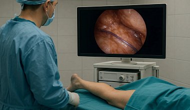
Endoscopic subfascial dissection of perforating veins in combination with traditional methods of surgical treatment of chronic venous insufficiency
Kazan State Medical Academy D.A. Slavin, L.E. Slavin, I.V. Fedorov, A.N. Chugunov, E.A. Shchedova The most severe complication of chronic venous insufficiency (CVI) are trophic ulcers. According to modern statistics, diseases of the veins of the lower extremities are an extremely common pathology. 20-25% of the population of industrialized countries suffer from chronic venous insufficiency, 1-4% have trophic changes in the skin. With conservative therapy, in 30% of cases of complete healing of ulcers, a relapse occurs within the first year [11]. In Russia, more than 35 million people suffer from various forms of CVI, and 15% of them already have trophic ulcers of the lower extremities. Chronic venous insufficiency is a pathological condition that results from two main causes: varicose veins and post-thrombophlebitic disease [3, 5]. Etiology and pathogenesis of varicose veins. Varicose veins of the superficial veins of the lower extremities is a polyetiological disease. To date, a number of endogenous and environmental factors playing a significant role in the origin of this pathology have been well studied [3, 5]. Risk factors: - Age. - Gender. - Pregnancy. - Burdened heredity. - Obesity. - Professional factors. With increasing age, the risk of developing varicose veins increases, which is associated with atrophy of the contractile elements of the venous wall and a decrease in its tone in the elderly. The maximum prevalence falls on the age group from 55 to 80 years [11]. Today, it is known that varicose veins of the lower extremities occur much more often in women than in men in a ratio of approximately 3:1. This is due to the secretion of female sex hormones and its physiological changes during pregnancy and menstruation. Estrogens have a direct effect on the venous wall, causing a decrease in its tone and an increase in distensibility [2]. Modern genetic studies have shown that early and severe forms of varicose veins are associated with an autosomal dominant inheritance pattern [17]. 50% of patients suffering from CVI have an adverse family history. A number of authors believe that varicose veins are caused by a hereditary, progressive imbalance between the different types of collagen in the vein wall [19]. Obesity also significantly increases the likelihood of developing varicose veins. A 20% increase in body weight leads to a fivefold increase in the risk of this pathology in women [10]. Varicose veins are most often found in individuals whose work requires them to remain in an upright position for long periods of time and who are subject to heavy physical exertion (surgeons, hairdressers, salespeople, loaders, etc.). Pathogenesis of varicose veins. The main hemodynamic factor leading to hypertension in the superficial venous system is blood shunting from the deep veins, which is associated with insufficiency of the valves at the mouth of the saphenous veins (vertical reflux) and incompetence of the perforators (horizontal reflux). Moreover, 90% of the latter is localized in the calf region, and 87% of them relate to the veins of Cockett I-III [3]. The severity of hemodynamic and trophic disturbances is determined not by the type of anastomosis, but by its level. The more distal the incompetent perforating vein, the greater the blood shunting pressure will be. Moreover, in addition to the high pressure caused by retrograde blood flow from the proximal parts of the superficial venous system, there is even higher pressure from the deep system, which is caused by retrograde blood flow through the incompetent perforators. Chronically elevated venous pressure, most pronounced in the distal parts of the leg, leads to tissue hypoxia and edema. As a result, severe hemodynamic and trophic disorders develop precisely in the lower third of the leg with Cockett perforating vein insufficiency [3, 5]. According to modern concepts, the key moments in the pathogenesis of trophic changes in CVI are dysfunction of the venous endothelium with subsequent disturbances in the venous wall-blood system, inflammatory processes involving activated leukocytes, and microcirculation disorders [9]. The result of inflammation is increased capillary permeability, the development of edema, endothelial damage by oxygen free radicals, aggregation and adhesion of neutrophils and platelets, and the release of coagulation factors with the formation of platelet clots. In addition, the rheological properties of the blood change. Red blood cells form "route columns" (sludge), plasma fibrinogen levels increase, fibrinolysis decreases, which leads to increased blood viscosity and a tendency to thrombus formation. Thus, chronic venous hypertension triggers a chain of pathological reactions that cause trophic changes in the skin of the lower extremities [8, 12]. International Clinical Classification of Chronic Venous Insufficiency CEAP: - Stage 0 No symptoms of venous disease during examination and palpation. - Stage 1 Telangiectasias or reticular veins. - Stage 2 Varicose veins. - Stage 3 Edema. - Stage 4 Skin changes due to venous disease: hyperpigmentation, venous eczema, lipodermatosclerosis. - Stage 5 Skin changes as mentioned above and signs of a healed ulcer. - Stage 6 Skin changes mentioned above and open ulcer. Types of surgical interventions: - Traditional surgeries. - Sclerosurgery. - Endosurgery. Basic principles of surgical treatment of varicose veins: - Elimination of pathological reflux from the deep to the superficial venous system. - Elimination of varicose veins. An important link in the pathogenesis of trophic disorders in CVI is the insufficiency of the perforating veins of the leg. Thus, the elimination of pathological horizontal reflux is the main method of treatment and prevention of trophic ulcers. For this purpose, various techniques have been proposed, in particular, the Linton, Felder, Cockett, and Sherman operations. One of the most effective interventions in the history of phlebology was the Linton procedure, developed by him back in 1938. It involved subfascial ligation of perforating veins through a wide incision extending almost the entire length of the leg. However, this technique is highly invasive, requiring a long hospitalization and rehabilitation period. The incision being performed in an area of ??trophic changes and a high rate of local complications ultimately lead to poor cosmetic results [1, 5, 16]. Subsequently, a technique of "blind" dissection of perforating veins was proposed. This involves inserting long scissors under the fascia through a small incision (3 cm) in the middle third of the leg, and blindly dividing any veins encountered by advancing them distally with open blades. This intervention is associated with massive bleeding, a high rate of local complications, and is not considered radical. The rapid development of endoscopic and minimally invasive surgery in the last 5-10 years has allowed to solve many problems that existed in surgical phlebology. In 1985, Hauer first published a work devoted to the endoscopic method of treatment of CVI. As a result, endoscopic subfascial dissection of perforating veins (ESPD) was developed and implemented, allowing to cross-section of all incompetent perforating veins under direct visual control and with minimal trauma, which is the key to effective treatment and prevention of varicose veins, its recurrence and complications [1, 2, 16]. It should be emphasized that this intervention is indicated for patients with trophic ulcers, which are a contraindication for traditional methods of surgical treatment of CVI. Advantages of endoscopic subfascial dissection of perforating veins: - Pathogenetic justification and radicality of the operation. - Minimal surgical trauma. - Low incidence of complications and relapses. - Good cosmetic result. - Reducing the duration of hospitalization and patient rehabilitation. In conclusion, it should be noted that currently, no single surgical method meets all the pathogenetic principles of CVI treatment, making a combination of them essential. Combinations of these or other surgeries primarily depend on the severity of pathological changes in the venous system of the lower extremities. Materials and Methods: Since October 2003, 17 endoscopic subfascial dissections of perforating veins (4 men and 13 women) have been performed at the surgical department of the Kazan Railway Hospital for chronic venous insufficiency associated with varicose veins. The average age of the patients was 57 years (range, 33 to 78 years). Disease duration ranged from 1 to 40 years. Preoperative diagnostics included inspection and palpation, as well as color duplex sonography of the venous system of the lower extremities with marking of incompetent perforating veins (Vivid 3 Expert, General Electric, USA). In 13 cases, ESPV was performed against the background of trophic changes in the skin (CEAP 4-6), of which 7 patients had open ulcers. Two patients were operated on for recurrent varicose veins after traditional interventions on the subcutaneous veins. All operations were performed under epidural anesthesia. Patients with trophic disorders received antibiotic prophylaxis (Cefotaxime 1 g intravenously, immediately before surgery). In the presence of at least one risk factor for thromboembolic complications associated with the patient's condition, low molecular weight heparin (fraxiparin (2850 IU anti-Xa) 0.3 ml subcutaneously once) and elastic compression were prescribed preoperatively. In 15 cases, a combined intervention was performed: crossectomy, ESPV, removal of the great saphenous vein and its varicose tributaries. Indications for ESDV Insufficiency of two or more perforating veins Combination of insufficiency of perforating veins with CVI due to varicose veins (CEAP 4-6) Trophic ulcer of the lower extremities Contraindications Arterial insufficiency of the lower extremities Postthrombophlebitic disease Acute inflammation Primary or secondary lymphedema Instruments and equipment required for ESDV Standard endovideosurgical stand Special endoscope with an instrument channel Sheath for advancement in the subfascial space Pneumatic cuff and fixator Dissector Bipolar forceps Clipper Scissors The use of a specially designed set of instruments and equipment is an important component of effective and safe endoscopic subfascial dissection of perforating veins. Surgical technique and prevention of complications. The first important stage of the combined surgery is the crossectomy, which involves isolating the saphenofemoral junction and high transection of the great saphenous vein and all its tributaries. For this purpose, in most cases, we used an inguinal approach of 3–4 cm in length, performed along the eponymous fold medial to the pulsation point of the femoral artery. After mobilizing the saphenofemoral junction, all tributaries of the great saphenous vein were isolated, ligated, and transected. The vein was then divided between clamps at a distance of approximately 1 cm from the orifice and ligated twice. The inguinal approach is associated with less trauma and a better cosmetic result compared to the oblique longitudinal approach [5]. To prevent recurrence, it is important to perform the crossectomy correctly, leaving no long stump or tributaries of the great saphenous vein. In addition, certain anatomical structures in this area must be completely intact. This is especially true for the femoral vessels and the external pudendal artery, which supplies the cavernous bodies in men. Endoscopic subfascial dissection of the perforating veins is the next stage of the surgery. To create graduated compression of the femoral vessels and ensure a dry surgical field in the subfascial space, a sterile cuff, pre-inflated to a pressure of 120-130 mmHg, was applied to the lower third of the thigh. A special fixator was used to prevent the cuff from slipping during surgery. To avoid ischemic complications, the duration of compression should not exceed 60 minutes. A 2-3 cm skin incision was made in the middle third of the leg along its medial surface, two fingerbreadths below the tibial margin. The subcutaneous tissue and fascia were dissected. Using a blunt finger, the fascia was dissected from the muscles. A sheath with an endoscope (Xion) was inserted into the subfascial space and explored. The endoscope is equipped with 300 optics and a working channel for inserting instruments of varying diameters (5 and 10 mm). The 230 mm sheath, with a dissecting olive at the end for advancement in the subfascial space, has a channel for smoke aspiration during coagulation. We did not use insufflation, as the unique design of the endoscope and sheath provides the necessary working space without gas supply, which in turn reduces surgical invasiveness and the risk of postoperative complications. It should be noted that, in an effort to identify all perforating veins during exploration, we did not limit ourselves to searching for veins detected by ultrasound. Clinical signs of their incompetence include an increase in vessel diameter (d > 3 mm) and the diameter of the openings in the fascia. Using a dissector, they were isolated from the surrounding tissue and, depending on their caliber, coagulated with bipolar forceps or clipped, and then divided with scissors. In this way, all perforating veins were sequentially processed. In most cases, we divided 3-4 incompetent veins. The use of bipolar coagulation ensures reliable hemostasis with a vessel diameter of up to 5-6 mm. If a larger vein is detected, a clip applicator must be used [2, 17]. A disadvantage of clipping is leaving foreign bodies in the subfascial space. At the end of this stage of the surgery, a revision of the subfascial space was performed to monitor hemostasis. After this, the endoscope was removed and the cuff was removed. The duration of the endoscopic procedure did not exceed 50 minutes. The next step was to make a small incision (1 cm) above the medial malleolus. The great saphenous vein was isolated. The distal portion was ligated, and a plastic phlebextractor was inserted into the proximal portion and advanced upward toward the inguinal region. Varicose veins in the saphenous veins were then isolated and removed through separate small incisions (1–2 cm). Surgical approaches, except for the inguinal one, were sutured, and aseptic dressings were applied. No drainage was performed, and the fascia was not sutured. The great saphenous vein was removed retrogradely through the inguinal incision using a phlebextractor. With this direction of saphenectomy, the risk of damaging the saphenous nerves and lymphatic collectors is minimal [5]. The inguinal approach was sutured last. An important detail of the operation is elastic bandaging of the limb up to the inguinal fold before removal of the great saphenous vein, which helps to avoid extensive hematomas in the postoperative period. Results and discussion. In the overwhelming majority of cases, the postoperative period was uneventful. All patients received Detralex 500 mg twice daily for two months, while elastic compression of the operated limb was applied. Eight weeks after surgery, trophic ulcers had healed in all patients. Skin grafting was not used; local treatment was primarily with Curiosin solution. No recurrences were observed. In two cases, we observed superficial infection in the postoperative wound area on the lower leg in patients operated on for trophic ulcers, which required antibiotics. Antibacterial therapy was not administered to the remaining patients. Sensory disturbance in the saphenous nerve area was observed in one patient. It is currently unclear whether this complication was related to the endoscopic stage of the surgery or to the subsequent removal of the saphenous veins. Hematomas along the great saphenous vein, which often accompany saphenectomy, were observed in seven patients. При этом необходимо отметить, что обширных подкожных и субфасциальных гематом мы не отмечали. Повторного вмешательства не потребовалось ни в одном случае. Такие осложнения, как тяжелая раневая инфекция, тромбоэмболия легочной артерии и тромбозы глубоких вен нижних конечностей, не наблюдались. Полученные нами результаты сопоставимы с литературными данными [13, 15]. Необходимость профилактики тромбоэмболических осложнений низкомолекулярными гепаринами обусловлена тем, что у больных с ХВН, особенно с трофическими расстройствами, имеются все предпосылки к образованию тромбов — повреждение эндотелия, застой и нарушение реологических свойств крови. Таким образом, хроническая венозная недостаточность повышает риск тромбоза как поверхностных, так и глубоких вен нижних конечностей. Дополнительная травматизация поверхностной венозной системы и перфорантов во время операции, наложение компрессионной манжеты, манипуляции в субфасциальном пространстве усугубляют ситуацию. Особенно при наличии у пациента других факторов риска. По литературным данным, частота венозных тромбозов нижних конечностей при операциях на поверхностных и перфорантных венах составляет 6 — 10%, частота тромбоэмболии легочной артерии (ТЭЛА) — 0,75%, летальность при которой достигает 30% [4, 7, 16, 18]. Примерно в 80% случаев венозные тромбоэмболические осложнения протекают бессимптомно, без клинических проявлений и обнаруживаются впоследствии при развитии ТЭЛА или хронической венозной недостаточности. При этом у 92% пациентов с доказанной тромбоэмболией был выявлен лишь один клинический фактор риска [6, 14]. Венозные тромбозы нижних конечностей также могут приводить к посттромбофлебитической болезни и рецидиву ХВН. В связи с этим считаем обязательным при наличии хотя бы одного из факторов риска тромбоэмболических осложнений, связанных с состоянием больного, проводить непосредственно перед вмешательством профилактику низкомолекулярными гепаринами. Нами отмечено, что введение фраксипарина (0,3 мл п/к однократно) не оказывает негативного влияния на ход операции. Таким образом, для предотвращения различных осложнений необходимо проводить профилактические меры, детально выполнять все рекомендации по технике операции и использовать соответствующее оборудование и инструменты. Небольшие сроки и количество наблюдений пока не позволяют делать нам окончательные выводы. Однако, несмотря на то, что эндоскопическая субфасциальная диссекция перфорантных вен является достаточно дорогостоящей и относительно трудоемкой процедурой, ее эффективность, патогенетическая обоснованность и экономическая целесообразность в сочетании с операциями на подкожных венах очевидны. Все вышесказанное свидетельствует о несомненных перспективах дальнейшего развития и широкого внедрения данного метода в клиническую практику в России. Литература: Егиев В.Н. Хирургия «малых пространств.»- М.: Медпрактика-М, 2002. — 56с. Кияшко В.А. Консервативное лечение хронической венозной недостаточности // Русский медицинский журнал. — 2002. — № 26. — С.1214-1219. Савельев В.С., Думпе Э.П., Яблоков Е.Г. Болезни магистральных вен. — М.: Медицина, 1972. — 440с. Савельев В.С. Послеоперационные венозные тромбоэмболические осложнения: фатальная неизбежность или контролируемая опасность? // Хирургия. — 1999. — № 6. — C.60-63. Яблоков Е.Г., Кириенко А.И., Богачев В.Ю. Хроническая венозная недостаточность. — М.: Издательство «Берег», 1999. — 128с. Bergqvist D. Optimal management of deep vein thrombosis // Vascular Surgery Highlights. — 2000. — P.39-46. Campbell B. Thrombosis, phlebitis and varicose veins // BMJ. — 1996. — № 312. — P.198-199. Dormandy J. Pathophysiology of venous leg ulceration // Angiology. — 1997. — № 48. — P. 71 — 75. Granger D., Schmid-Shoenbein G. Chronic venous ulceration: a role for leukocyte-mediated injury. In: Loosemoore T., Dormandy J. Physiology and Pathophysiology of Leukocyte Adhesion. — USA: Oxford University Press: 1995. — P. 447-457. Jantet G. Эпидемиолгия заболеваний вен // Int. Angiol. — 1997. — № 163. — P.6-8. Mayer W., Jochmann W., Partsh.H. Varicose ulcer: healing in conservative therapy. A prostpective study // Wein-Med. Wochenschr. — 1994. — № 144. — Р.250-252. McEver R. Leukocyte-endothelial cell interactions // Curr. Opin. Cell. Biol. — 1992. — P.4840-4849. Nelzen O. Prospective study of safety, patient satisfaction and leg ulcer healing following saphenous and subfascial endoscopic perforator surgery // Br. J. Surg. — 2000. — № 87. — P.86-91. Pieralli E., Prisco D., Fredi S. Клинические и связанные с гемостазом факторы риска у пациентов с тромбоэмболией // Int. Angiol. — 1997. — № 163. — C.12-13. Pierik EG, Van Urk H, Hop WC, Wittens CH Endoscopic versus open subfascial division of incompetent perforating veins in the treatment of venous leg ulceration: a randomized trial // J. Vasc. Surg. — 1997. — № 26. — P.1049-1054. Pietravallo A., Pataro E., Cocozza C., Vidal R., Dandolo M. Эндоскопические вмешательства при недостаточности перфорантных вен нижних конечностей // Флеболимфология. — 2003. — № 19. — C.5-12. Pistorius M. Chronic Venous Insufficiency // Angiology. — 2003. — Vol.54. — suppl. 1. — Р.5-12. Samama M. Хроническая венозная недостаточность, варикозное расширение вен и тромбоз глубоких вен нижних конечностей // Медикография. — 2000. — № 3. — C.59-62. Sansilverti-Morel P., Rupin A., Badier-Commander C., Fabiani J., Verbeuren T. Chronic venous insufficiency: Dysregulation of Collagen Synthesis // Angiology. — 2003. — Vol.54. — suppl. 1. — P.13-18.


