

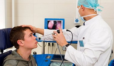
Endoscopic examination in the diagnosis of sphenoid sinus diseases
A.M. Ashurov Tashkent Institute of Advanced Medical Studies Tashkent, Uzbekistan Relevance of the problem. The problem of sphenoiditis has been and remains relevant in rhinology, especially in countries with a hot climate (Central Asia). Currently, the leading diagnostic methods for sphenoiditis are computed tomography and magnetic resonance imaging (CT and MRI). The results of CT and MRI in the diagnosis of sinusitis cannot be 100% trusted, since CT and MRI often exhibit overdiagnosis (Yu.M. Ovchinnikov, 1997). Objective of the study: To identify endoscopic signs of sphenoid sinus (SS) inflammation. Materials and methods. We observed 40 patients (22 men, 18 women) aged from 16 to 60 years. The diagnosis of sphenoiditis was made based on the patient's complaints, disease history, endoscopic examination, CT and MRI results. A rigid endoscope with a diameter of 4 mm and end and side optics with visual angles of 0° and 30° was used for endoscopy. During endoscopic examination, abnormalities in the architecture of the nasal cavity were revealed in all patients: a deviated nasal septum, hypertrophy of the middle turbinate, medial protrusion of Onodi cells, adhesions in the area of ??the natural ostium of the superior nasal passage, polypous ethmoiditis, etc. The endoscopic picture in acute sphenoiditis was accompanied by hyperemia and edema of the mucous membrane of the superior nasal meatus, around the natural ostium of the superior nasal passage; discharge from the superior nasal passage was noted in exudative inflammation. Chronic sphenoiditis was accompanied by hyperplasia of the mucous membrane around the natural ostium and thick secretion. Conclusions: Endoscopic signs of sphenoiditis are: changes in the mucous membrane of the middle nasal meatus, around the natural ostium of the superior nasal passage, and the presence of discharge.


