

New fluid management system for hysteroscopic surgeries
23.01.2017 16:25:00
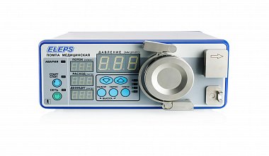
I.V. Klyucharov, A.A. Khasanov, I.R. Galimova, R.N. Farkhutdinov Journal "Practical Medicine" No. 4, August 2016 / Volume 2 A New Fluid Management System for Hysteroscopic Surgeries Many technical issues and complexities associated with operative hysteroscopy and hysteroresection are associated with the management of the fluid medium used for uterine cavity distension. For the operating room nurse, this involves monitoring the moments when the reservoir with the supplied fluid is empty (a new one must be connected) and the overflow of the fluid collection container (it must be emptied), as well as the task of constantly calculating the deficit of the used fluid. The latter is especially difficult, as there are many factors that distort the data. Fluid reservoirs can be "underfilled" or "overfilled" by up to 10%; in addition, it is necessary to account for the fluid remaining in them, as well as fluid on the floor and surgical linens. For the surgeon, the primary concern is achieving good visualization of the uterine cavity, which depends on the pressure in the uterine cavity and the rate of fluid delivery. Both the surgeon and the patient are aware of and concerned about potential complications arising from excessive absorption of the fluid used to distend the uterine cavity and the subsequent development of life-threatening hyponatremia and hypervolemia. The device we developed addresses these concerns. Key words: fluid flow management, hystero pump, complications, fluid balance (deficit), hypervolemia, hyponatremia. Surgical hysteroscopy is a commonly used gynecological surgical procedure for the treatment of various intrauterine pathologies. During resectoscopic surgeries, both arterial and venous vessels of the endometrium and myometrium are often exposed, leading to intravasation of the fluid dilating medium. When using 0.9% saline as a dilating medium during surgery, electrolyte disturbances are not severe. At the same time, when using non-electrolyte solutions such as glucose, glycine, and sorbitol, the risks of electrolyte disturbances leading to hypervolemia and hyponatremia increase dramatically. Often, due to the inability to accurately monitor the balance between injected and excreted fluid, the surgeon is unaware of the impending catastrophe. The essence of the problem: The surgeon performing a hysteroscopic procedure must ensure good visualization—a clear field of view. To achieve this, the uterine cavity must be sufficiently dilated for viewing (intrauterine cavity pressure), and the rate of fluid exchange must be sufficient to ensure the removal of tissue debris, clots, and liquid blood that could contaminate the field of view (fluid flow rate). There are several methods for delivering the dilating medium into the uterine cavity. A gravity-fed system, with or without an auxiliary cuff, delivers fluid at a pressure and velocity dependent on the difference in height between the solution container and the operating table, the cuff pressure, the diameter and material properties of the fluid inlet and outlet tubes, the design of the hysteroscope's internal channels, the scope of the hysteroscope's coverage of the cervix, and the degree of opening of the outflow stopcock. Considering the influence of these factors significantly complicates intrauterine hysteroscopic interventions. It is also important to note that, compared to laparoscopy, hysteroscopy is not a closed system, but rather a system with a constant pressure gradient that decreases from the source to the uterus and outflow tract. Therefore, true pressure can only be determined using a separate sensor placed in the uterine cavity. When the outflow tract is closed, the pressure in the uterine cavity can be maximal; however, a weak flow can impair visualization, while uterine distension is maximal. Conversely, with a fully open port, the uterine cavity can be effectively cleared of contaminants, but the pressure in the uterine cavity may be insufficient. Fluid Flow Management System (FMS) The Advanced Fluid Management System (AFMS) was designed to ensure increased patient safety and provide key features necessary for hysteroscopic surgery: high accuracy in matching the set pressure to the actual pressure in the uterine cavity, precise accounting of the balance of used and returned fluid, and the ability to use a variety of fluid containers. Hysteropump: The heart of the system is the hysteropump, whose primary function is to precisely maintain the pressure and flow rate of fluid into the uterine cavity to support hysteroscopic procedures (Fig. 1). [ /upload/medialibrary/efa/1.jpg ] Fluid is supplied from three possible sources: 400-500 ml bottles of sterile liquid installed in a suitable bracket, bags of sterile liquid suspended from a suitable holder, and sterile jars from which any liquid sterile solution can be dispensed. The solution is supplied to the hysteroscope's inlet tract using a peristaltic pump and silicone tubing of a specified diameter and length. In this hystero pump, two elements are crucial in the algorithm for maintaining the required pressure: consideration of the effect of the hysteroscope's design (hydrodynamic profile) on the pressure in the uterine cavity and compensation for the pressure drop when the outflow stopcock is opened. These two key features of the hysteropump operation help minimize the previously identified experimental error introduced when using sets of devices and instruments from different manufacturers (see table) and the distortion manifested in a significant drop in pressure in the uterine cavity when opening the outflow tap during surgery (Fig. 2) or using a system with automatic evacuation of the contents of the uterine cavity [1]. The use of these functions allows for resectoscopy operations with minimal pressure parameters in the uterine cavity (50-70 mmHg). The most important element of the safety of hysteroscopic operations is ensuring the calculation of the deficit of the used fluid. There are two methods for calculating the fluid balance: volumetric - based on calculating the volume of the used fluid, and gravimetric - based on calculating the weight of the used fluid. Currently, the results of a study of balance (deficit) calculation errors have been published, which are associated with permitted tolerances in the production of containers with liquid within ±10% of the declared volume [2]. For example, if we used 10 liter bags of fluid during surgery, a 10% underfill would result in up to 1.0 liter of fluid being erroneously accounted for, while a 10% overfill would result in up to 1.0 liter being erroneously omitted when calculating the fluid balance (deficit). It's important to remember that current recommendations call for terminating the surgery if the deficit is 0.5 liters of non-electrolyte solution or 2.0 liters of electrolyte solution, due to the risk to the patient's health and life [3]. The essence of the gravimetric system implemented in the PSUZH is that the fluid supply and return containers are located on electronic scales, and the system continuously weighs the amount of fluid in the containers on the scale. The amount of fluid not returned to the scale constitutes the fluid balance (deficit), which is continuously calculated during the surgery and reported in the final report. A key element of the system is ensuring the continuity of fluid supply and withdrawal during surgery. The electronic unit of the hysteropump stores the fluid balance (deficit) value when replacing the containers from which the sterile medium is dispensed and into which it is collected after use. To replace the fluid, it is necessary to stop the pump, change the container, and, after restarting the pump, continue the operation. This ensures a minimal delay in the operation. For quick replacement, the set of containers includes 3 jars (3x1.8 l) for sterile solution and 3 jars (3x1.8 l) for collecting used fluid. Discussion The fact that patients can suffer as a result of hysteroresectoscopy surgery is the best evidence for the need to use an automated fluid delivery system with an integrated gravimetric balance (deficit) control system [4]. If 60 mmHg is sufficient to distend the uterine cavity and perform the operation, then why use higher parameters? If bleeding occurs during surgery, increasing the intrauterine pressure to the level of mean arterial pressure in most cases allows for improved visibility and continuation of the operation. This increase will not be accompanied by significant intravasation of fluid into the vascular bed and, accordingly, will not increase the risk of developing such serious complications as vascular bed fluid overload (hypervolemia) and hyponatremia. This is convincingly demonstrated by the experience of colleagues [5] and our own experience. Increasing the fluid flow rate is another option for solving the problem, but intravasation is proportional to the flow rate [6]. Since intrauterine pressure is the only important factor causing intravasation, accurate pressure measurement and the ability to change it are of fundamental importance. The PSUG described in this article allows the operator to accurately preset and change the pressure of the medium intraoperatively to ensure adequate visualization with minimal pressure in the uterine cavity. The use of PSUG allows for much more accurate control of the pressure and flow of fluid in the uterine cavity compared to many hysteropumps that do not have the following capabilities: 1. Selection of the hydrodynamic profile of the hysteroscope used; 2. An algorithm for compensating for pressure changes when opening the outflow tap; 3. Continuous automatic monitoring of the fluid balance (deficit); 4. The presence of voice prompts on the amount of fluid used, every 500 ml of deficit and in the event of a sharp imbalance of fluid (for example, in case of uterine perforation). The advantage over systems delivering fluid by gravity, using a cuff and a large syringe is undeniable, since these systems in principle do not allow for precise control of the fluid supply parameters [7]. The fluid flow can be regulated by changing the degree of opening of the hysteroscope outflow tap, while the decrease in pressure in the cavity will be insignificant, since the designed pump has an algorithm for compensating for maintaining the set pressure (Fig. 3). An additional advantage of using the PSUZH is that the nurse is freed from constant monitoring of the amount of supplied and returned fluid, as well as from monitoring the emptying of the reservoir with the supplied fluid, overflow of the reservoir with the returned fluid and the timeliness of their replacement, which helps reduce the workload of the operating room staff. The operating room staff receives the necessary signals in the form of voice prompts. [ /upload/medialibrary/5a8/3.jpg ] Conclusion Very often, gynecologists and nurses in the operating room, focused on the technical performance of a hysteroscopic surgery, do not notice significant intravasation, which can occur before the fluid reaches the limit of deficit. This system was created to ensure the primary task - patient safety. At the same time, it ensures comfortable work for the surgeon and the operating team as a whole, possessing high precision and simple and intuitive operation. With the use of the PSUZ, the risks of water-electrolyte imbalance when using saline are virtually reduced to zero, and the risks associated with the use of dielectric solutions are significantly reduced. More than 100 operations have been performed using this PSUZ in various clinics of the Russian Federation (10 were performed at the Republican Clinical Hospital of the Ministry of Health of the Republic of Tatarstan). In not a single case have there been any intra- or postoperative complications associated with the regulation of fluid flow. The system correctly took into account the fluid deficit during the operation and provided predictable working conditions for the operating team. [ /upload/medialibrary/34f/2.jpg ] REFERENCES 1. Klyucharov I.V., Prokhorov E.I., Borisov S.L., Kadyrov R.F. et al. Preset and actual pressure in the uterine cavity. Accuracy of our hysteropumps // Practical Medicine. - 2015. - No. 4 (89), Vol. 1. - P. 79-82. 2. Nicolopoulos I., Phillips G. Reliability of fluid monitoring during operative hysteroscopy // Gynecological surgery. - 2016. - Vol. 13 (1). - P. 23-26. 3. Hysteroscopic fluid monitoring guidelines // J. Am. Assoc. Gynecol. Laparosc. - 2000. - 7. - P. 167-168. 4. Rademaker B.M.P., van Kesteren P.J.M., de Haan P. et al. How is intravasation limit in hysteroscopic surgery? //JMIG. - 2011. - Vol. 18(3). — P. 355-361. 5. Garry R., Hasham F., Kokri M.S. et al. The effect of pressure on fluid absorption during endometrial ablation // J. Gynecol. Surg. - 1992. - 8. - P. 1-10. 6. Corson S.L., Brooks P.G., Serden S.P. et al. Effects of vasopressin administration during hysteroscopic surgery // J. Reprod. Med. - 1994. - 39. - P. 419-423. 7. Wan-Hua Ting, Ho-Hsiung Lin, Ming-Ping Wu et al. Safety and efficacy of manual syringe infusion of distending media for hysteroscopic procedures: a case-control study // European journal of obstetrics and Gynecology and reproductive biology. — 2015. —191. —R.112-115.


