

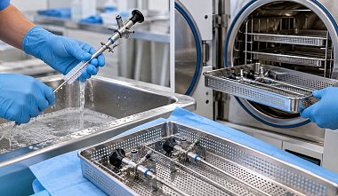
Hysteroscopes are used in gynecology to examine the uterine cavity, diagnose pathological changes, and perform intrauterine procedures. After each procedure, the equipment undergoes an established cleaning, disinfection, and sterilization cycle. When preparing it for reuse, it is necessary to preserve the accuracy of the optical system, the integrity of the working components, and the reliability of the connections.
Sterilization of a hysteroscope in an autoclave is permitted only when explicitly specified by the manufacturer. High temperature and pressure are not suitable for every model or accessory. Therefore, when equipping a gynecology department, it is important to consider in advance the permitted reprocessing methods, optical characteristics, and equipment maintenance requirements.
Why Hysteroscopes Require Thorough Cleaning and Sterilization
A hysteroscope is inserted through the cervical canal into the uterine cavity to examine the mucous membrane and perform diagnostic or surgical procedures. During the procedure, blood, mucus, and tissue particles remain on the optical tube, sheath, and working instruments. If these contaminants dry, they become more difficult to remove. Residual biological material may remain at component joints, in recesses, and in channels, where the design includes them.
Cleaning removes contaminants, disinfection reduces the number of microorganisms, and sterilization destroys all forms of microorganisms, including spores. These stages cannot replace one another. Processing an instrument that has not been cleaned reduces the effectiveness of subsequent procedures: organic residues prevent the sterilizing agent from contacting the surface of the device.
The preparation procedure depends on the endoscope’s design and accessories, the intended use of the medical device, and the manufacturer’s requirements. Rigid optics, sheaths, stopcocks, and working components are inspected and processed according to their design. Instructions for one endoscope must not automatically be applied to another model, even if both instruments are used in gynecology.
SanPiN 3.3686-21, as amended on June 25, 2025, establishes sanitary requirements for the prevention of infectious diseases, including the processing of equipment used for endoscopic procedures. The device’s operating instructions must also be followed. Steam sterilization in an autoclave is performed only when expressly permitted by the manufacturer. If the material, optical assembly, or an individual component is not designed to withstand high temperature and pressure, an unsuitable cycle may damage the equipment.
Steps for Processing a Hysteroscope Before Sterilization
Immediately after hysteroscopy, visible traces of blood, mucus, and tissue are removed from the equipment. Contaminants must not be allowed to dry on the optical tube, sheath, or working instruments: dried residues are more difficult to remove from surfaces and hard-to-reach areas. Cleaning and disinfecting agents compatible with the endoscope materials are used.
-
Preliminary cleaning. External surfaces are wiped with a soft, lint-free cloth to remove visible contaminants. Removable components are cleaned separately. If the design includes channels that can be flushed, the solution is passed through them using suitable adapters. Abrasive sponges and stiff brushes may scratch the optics and damage coatings, so they must not be used.
-
Disassembly. The sheath, stopcocks, valves, and working instruments are disconnected if the design allows them to be disassembled. Each component is cleaned separately, paying particular attention to threads, joints, and recesses where biological contaminants may remain. The optical tube must not be disassembled: its internal components require specialized servicing.
-
Cleaning and disinfection. Components are processed manually or in a washer-disinfector suitable for the equipment. During manual cleaning, joints and hard-to-reach areas are treated thoroughly. The solution concentration, temperature, and contact time must comply with the instructions for the product being used.
-
Rinsing. Residual cleaning and disinfecting solutions are removed from surfaces and channels after processing. Inadequate rinsing may leave chemicals on the components, potentially damaging materials or affecting subsequent processing. Water of the required quality is used for rinsing.
-
Drying. Moisture is removed from external surfaces and from channels, if present. No droplets or streaks should remain on the optics that could interfere with inspection. The drying method is selected according to the device’s design: unsuitable temperature or air pressure may damage individual components.
After these steps, the hysteroscope is inspected. If any contamination remains on the surface, cleaning is repeated. Only a clean, dry, and serviceable device may be prepared for sterilization.
Cleaning, disinfection, and sterilization serve different purposes. Cleaning removes blood, mucus, and other contaminants; disinfection reduces the number of microorganisms; sterilization destroys viable microorganisms, including spores. Loading a contaminated hysteroscope into an autoclave does not replace cleaning and does not guarantee successful sterilization.
Inspecting the Hysteroscope Before Sterilization
Before loading the sterilizer, confirm that the equipment is clean, in working order, and compatible with the selected processing method. Damage to the optics, body, or connections can impair visualization, compromise the integrity of channels designed to be sealed, and cause the device to fail during a procedure. Inspection must be performed by trained personnel in accordance with the manufacturer’s instructions. Check the following components:
- Optical tube and distal lens. There must be no visible contamination, chips, cracks, or other damage on the surface.
- Eyepiece or video-system connection. Check the integrity of the connections and look for signs of damage.
- Body and adapters. Inspect the connection points for the light guide and other components.
- Sheaths, stopcocks, and working components. Check the condition of the parts, the movement of any mechanisms provided, and the integrity of the seals.
- Channels and connections. If these are part of the design, check their patency and condition using the method specified by the manufacturer.
If defects are found, the device must not be returned to use until the cause has been investigated and a decision has been made regarding repair or replacement. Do not attempt to repair optical defects by mechanical treatment or use products not intended for the relevant material.
The labeling and documentation must also be checked. They should specify the permitted sterilization method and any necessary restrictions. If compatibility with steam sterilization has not been confirmed, do not autoclave the device.
Preparing a Hysteroscope for Autoclave Sterilization
Before deciding how to sterilize a hysteroscope in an autoclave, check the operating instructions for the specific model. They must specify the permitted processing method and restrictions regarding temperature, pressure, exposure time, packaging, and placement in the sterilizer. If steam sterilization is not approved, the autoclave must not be used.
Preparation includes several steps:
- Confirm that the device is approved for saturated-steam sterilization.
- Make sure cleaning, rinsing, and drying have been completed.
- Disassemble removable components in accordance with the instructions.
- Set stopcocks and other components to the positions specified by the manufacturer.
- Package the device in material compatible with the selected cycle and approved for sterilizing medical devices.
- Position the optics and accessories so that they do not touch heavy instruments or become subject to impact or pressure.
Before loading, check the autoclave itself: its technical condition, the selected program, the suitability of the packaging, and the permitted chamber load. Do not load items more densely than the sterilizer’s instructions allow. Overloading and incorrect positioning prevent proper processing. If the manufacturer does not specify whether autoclaving is permitted, or if the equipment documentation has been lost, do not select a cycle by analogy with another hysteroscope. Obtain official information about the permitted processing method for the device. The specific model and its configuration matter—not merely the instrument’s name or the material of its body.
How Hysteroscope Sterilization in an Autoclave Works
Steam sterilization uses saturated water vapor under pressure. The steam transfers heat to the instrument’s surfaces and, when the specified parameters are maintained, destroys viable microorganisms, including bacterial spores. The outcome depends on temperature, exposure time, and adequate steam contact with the surfaces being processed.
At the beginning of the cycle, air is removed from the autoclave chamber and, where the sterilizer’s design provides for it, from the cavities within the load. Air can prevent steam penetration and uniform heating of the devices. The chamber is then filled with saturated steam, and the temperature rises to the set value. During the sterilization holding phase, the instrument is maintained at the specified temperature for a defined period. The combination of temperature, time, and steam exposure produces the sterilizing effect. After the holding phase, steam is removed from the chamber. Drying then takes place as specified by the sterilizer program: residual moisture is removed from the chamber and sterilization packaging. Dry packaging is necessary to preserve sterility during subsequent storage. If the packaging remains wet, its protective properties may be compromised.
The temperature and holding time must be selected according to the approved cycle for the specific hysteroscope model. There are no universal parameters for all endoscopic equipment. High temperature and pressure may damage lenses, bonded joints, seals, and other sensitive components if the device is not designed for steam processing. Therefore, autoclaving is permitted only with the manufacturer’s explicit approval.
Sterilization Quality Control
After the autoclave program has finished, confirm that the cycle was completed without errors and that the hysteroscope can be released for use. The staff member responsible checks the recorded sterilization parameters, any sterilizer messages, and the condition of the packaging.
The following must be assessed first:
- whether the temperature, pressure, and exposure time specified by the program were achieved;
- whether the cycle finished without errors or emergency interruptions;
- whether the packaging remains intact, dry, and sealed;
- whether the chemical indicators meet the established acceptance criteria;
- whether the cycle has been recorded in the logbook or electronic tracking system.
Physical parameters are assessed using the sterilizer’s records; chemical indicators are assessed by the change specified by their manufacturer. Biological monitoring is performed at the frequency established by the medical organization and in the circumstances required by applicable regulations. One type of monitoring does not replace the other checks.
A hysteroscope must not be released for use if the autoclave failed to reach the required parameters, the cycle was interrupted, the packaging is damaged or wet, or the indicator results do not meet the established criteria. The device must undergo reprocessing in accordance with the approved procedure. The cause of the failure must be identified before the next cycle, especially if a sterilizer malfunction is suspected.
Sterilization results must be recorded so that the date and cycle number, the autoclave used, the program parameters, and the monitoring results can be identified. If a failure is detected, these records help determine which devices were processed in the affected load and prevent their use until an appropriate decision has been made.
Storage of Sterile Hysteroscopes
The following rules must be observed during storage:
- Do not open the packaging until the device is needed.
- Check the integrity of the packaging and the presence of labeling before the device is sent to the operating room.
- Observe the established storage periods and conditions.
- Protect the optical tube from impacts, pressure, and contact with hard objects.
- Track sterile devices and monitor the movement of equipment between departments.
The period for which sterility is maintained depends on the packaging, sterilization method, storage conditions, and applicable requirements. A single storage period must not be assigned to all hysteroscopes without considering these factors. If the packaging becomes wet, torn, or loses its seal, the device must be reprocessed before use. After sterile packaging is opened, the instrument must be used in accordance with the medical organization’s rules. An unused hysteroscope may be returned to storage as sterile only in accordance with applicable requirements and the facility’s internal procedures.
Common Errors in Hysteroscope Processing and Sterilization
Errors at any stage may result in inadequate cleaning, damage to the optical system, or loss of sterility. The most common problems arise for the following reasons:
- Autoclaving without checking the instructions. Not all hysteroscopes can withstand exposure to saturated steam at elevated temperatures. Before selecting a program, confirm that the manufacturer permits steam sterilization of the specific model.
- Incomplete cleaning before sterilization. Blood and tissue residues may remain in joints and hard-to-reach areas. If contamination is found after cleaning, repeat the process.
- Use of incompatible chemical agents. Unsuitable products may damage optical surfaces, coatings, and seals. Select cleaning and disinfecting agents in accordance with the device instructions.
- Incorrect disassembly. Opening non-disassemblable assemblies or mishandling removable components may damage the instrument. Disassembly and reassembly must follow the manufacturer’s instructions.
- Careless loading of the autoclave. Contact between the optics and metal instruments, container walls, or other devices may cause scratches and chips. Secure the hysteroscope in a holder designed for it.
- Changing sterilization parameters. Independently adjusting temperature, pressure, or exposure time disrupts the established cycle and may damage the equipment. Select the program based on the documentation for both the device and the sterilizer.
- Using devices after an unsuccessful cycle. An autoclave error, unsatisfactory monitoring results, or damaged packaging requires investigation and reprocessing in accordance with the approved procedure.
- Failure to observe storage conditions. Wet, opened, or damaged packaging cannot ensure continued sterility. Check its condition before use.
To prevent these errors, a medical organization needs an approved processing protocol, staff training, and access to current instructions. Staff must know which methods are permitted for each hysteroscope model, how to verify sterilization results, and what to do if a malfunction or cycle failure is detected.
ElePS is a Russian manufacturer of endoscopic optics, medical equipment, and instruments for endoscopic surgery. Its product range includes hysteroscopes and cystoscopes with various diameters and viewing angles, including specific models designated as autoclavable, as well as sheaths and accessories for hysteroscopy. When selecting equipment for a gynecology department, it is necessary to consider the device’s intended use, component compatibility, and permitted processing methods. The company’s specialists can clarify product specifications and help select equipment suited to the needs of the medical facility.


