

Surgical thoracoscopy. Benign mediastinal tumors.
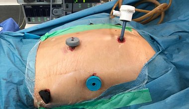
E.I. Sigal, R.G. Khamidullin Video-assisted thoracoscopy can be considered the method of choice in the treatment of benign tumors and cysts of the mediastinum, since the invasiveness of conventional thoracotomy in some cases is significantly higher than the invasiveness of the tumor removal stage itself. Even small mediastinal neoplasms force the surgeon to make disproportionately large incisions in the chest wall. Indications — Coelomic cysts of the pericardium. — Mediastinal lipomas. — Benign thymomas. — Thymic cysts. — Neurogenic tumors of the mediastinum. — Mediastinal teratomas. Mediastinal fibromas. Contraindications — General contraindications to TO. — Significant tumor size is a relative contraindication, since tumors larger than 10 cm in diameter can often be removed. Topographic anatomy of the mediastinum. Several schemes for dividing the mediastinum into floors and sections have been proposed. For video-assisted thoracoscopic surgery, the most common division is into five zones: anterosuperior, anteroinferior, posterosuperior, posteroinferior, and central. Thymomas are localized in the anterosuperior zone, coelomic pericardial cysts and mediastinal lipomas in the anteroinferior zone, and neurogenic tumors in the posterior mediastinum. Based on the above-described zones and the relatively constant location of tumors, there are clearly defined insertion points for thoracoports during mediastinal surgery. Surgical technique. The surgery is performed under general endotracheal anesthesia with separate ventilation of both bronchi. The first thoracoport for the thoracoscope is inserted into the 5th intercostal space along the anterior axillary line. After examining the chest organs and determining operability, instrumental thoracoports are inserted. Their number determines the nature of the tumor and the extent of the proposed surgery. Two thoracoports are usually sufficient. The insertion points for instruments depend on the tumor location. If the tumor is located in the anterosuperior zone of the mediastinum, instruments are inserted into the 5th intercostal space along the parasternal line and into the 3rd intercostal space along the posterior axillary line. For tumors in the anterior-inferior zone, trocars are inserted into the 3rd intercostal space along the anterior axillary line and into the 5th intercostal space along the posterior axillary line. For pathology in the posterosuperior zone, instrument insertion points are located in the 3rd intercostal space along the anterior axillary line and in the 5th intercostal space along the parasternal line. For tumors in the posteroinferior zone, trocars are inserted into the 3rd intercostal space along the parasternal line and into the 5th intercostal space along the posterior axillary line. For work in the central zone, instruments are positioned in the 3rd intercostal space along the anterior axillary line and in the 5th intercostal space along the posterior axillary line. The surgical technique is no different from that generally used in open surgery. After incising the mediastinal pleura, the tumor is isolated, the vessels feeding the tumor are dissected, then clipped or coagulated. If adhesions to the lung parenchyma are present, endoscopic suturing devices may be used. Large cysts are punctured and enucleated to facilitate manipulation. The cyst membranes are then excised. After complete enucleation of the tumor, the specimen is placed in a container and removed through the largest diameter thoracoport. For large tumors, a 4-6 cm mini-thoracotomy is performed to extract the tumor. At the end of the surgery, careful hemostasis and tumor bed exploration are performed. Under visual control, after drainage of the pleural cavity, the instruments are removed, and the wounds are sutured. Complications and their prevention. Given the anatomical features of the mediastinum, the complex relationships between vital organs, and the proximity of major blood vessels and the heart, rough manipulation and inexperienced surgeons can lead to damage, resulting in profuse bleeding, requiring urgent thoracotomy. Detailed visualization of all anatomical structures and precise surgical technique are the key measures to prevent serious intra- and postoperative complications.


