

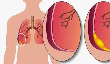
Surgical thoracoscopy. Pleural empyema.
E.I. Sigal, R.G. Khamidullin: If conservative therapy with punctures, closed drainage, and debridement of the pleural empyema cavity fails, thoracoscopy is used. Surgical technique. The procedure is performed under general anesthesia with artificial ventilation. A trocar for the thoracoscope is inserted into the pleural empyema cavity at a point determined by radiographic examination. The exudate is evacuated and sent for bacteriological examination. The pleural empyema cavity is then examined, and a thoracoport for instruments is inserted at the most convenient location. An electrocoagulation hook is used to separate the adhesions, thus creating a single cavity. Necrotic tissue and fibrin are removed with instruments or by aspiration. The pleural empyema cavity is sanitized with antiseptics, irradiated with a CO2 laser or ultraviolet irradiation, and then drained. Postoperative period. The empyema cavity is actively drained and treated with antiseptic solutions. Detoxification and potent antibacterial therapy are prescribed according to established protocols.


