

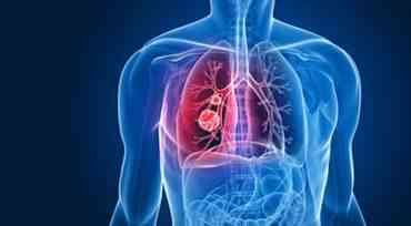
Surgical thoracoscopy. Peripheral lung tumors.
E.I. Sigal, R.G. Khamidullin. Differential diagnosis of peripheral benign and malignant lung neoplasms is complex. Peripheral lung tumors and solitary metastases are indications for surgical treatment. Despite indirect radiographic and tomographic signs of benignity, a definitive diagnosis is established only by histological examination of the specimen. Bronchoscopic examination is uninformative, especially with small peripheral tumors. Percutaneous needle biopsy for malignant neoplasms allows for diagnosis in 43-76% of cases; for benign neoplasms, the sensitivity of this method is significantly lower. Elderly patients with T1-2N0 peripheral lung cancer with low functional data due to severe comorbidity are indicated for atypical lung resection. The scope of intervention for benign peripheral tumors and solitary metastases is atypical lung resection or tumor enucleation. Indications for thoracoscopic surgery 1) Peripheral benign lung tumors. 2) Solitary pulmonary metastases. 3) Peripheral lung cancer T1-2N0 (in patients with LSN). Surgical technique. Thoracoscopy is performed under general endotracheal anesthesia with separate bronchial intubation. Lung ventilation is turned off on the side of the operation. A thoracoport for a thoracoscope is inserted into the 5th intercostal space along the midclavicular line. The pleural cavity and lung are examined. Thoracoports for instruments are inserted under visual control at the points most convenient for manipulation (the choice of insertion points for instrumental thoracoports depends on the location of the tumor in the lung). During the operation, 2-3 thoracoports are usually sufficient. Tumors located subpleurally are accessible for examination. Tumors located deep in the lung parenchyma are detected by one of the methods listed below. 1) Preoperatively, under CT guidance, a percutaneous puncture of the tumor is performed using a special needle with a hook at the end, and a methylene blue solution is injected. During thoracoscopy, the location of the tumor is determined by lung staining and the location of the needle. 2) Intraoperative ultrasound is performed using a special endoscopic transducer. Lung echolocation does not always allow determining the tumor location due to the airiness of the lung parenchyma. 3) Instrumental palpation of the lung. Using a dissector, the lung parenchyma is palpated, determining the location of the pathological formation. 4) Manual palpation of the lung. One of the punctures in the chest wall is widened, a finger is inserted into the pleural cavity, and the lung is palpated at the site of the suspected tumor location. This method of detecting peripheral tumors is the simplest and most effective. After detection, the tumor is punctured. The punctured material is subjected to urgent cytological examination. Depending on the cytological examination results, a specific surgery is performed. Atypical lung resection. A stapling device is applied to the lung tissue along the proposed resection line. The desired lung area containing the tumor is resected. Depending on the volume of the lung area to be removed, the device is applied up to 5-6 times. Lung tumor enucleation. The lung tissue is dissected over the tumor using electrosurgical instruments (scissors or a hook). The tumor is enucleated. The integrity of the lung is restored by applying endoscopic sutures, with the knots tied intra- or extracorporeally (Fig. 2). The removed tumor is subjected to urgent histological examination. If the process is confirmed to be benign, the surgery is terminated. The presence of a malignant tumor is an indication for anatomical lobectomy, performed thoracoscopically or conventionally. The pleural cavity is filled with fluid and the lung is gradually inflated to control pneumostasis: the appearance of air bubbles indicates insufficient tightness and requires the application of additional sutures to the lung. The lung is inflated after draining the pleural cavity, thoracoports are removed, and the surgical wounds are sutured. Anatomical lobectomy. Malignant peripheral lung tumors require anatomical lobectomy. Moreover, in lung cancer, a complete lymphadenectomy is important. The TO technique is similar to traditional lobectomy. Anatomical lobectomy is also used for bronchiectasis and chronic lung abscess. Indications: - Peripheral lung cancer T1-2N0. - Bronchiectasis in remission. - Chronic lung abscess. Contraindications: - Central lung cancer. - The presence of metastases in the root of the lung. - Fusion of the interlobar fissures. Surgical technique. The operation is performed using four thoracoports or supplemented by a mini-thoracotomy (in the 4th or 5th intercostal space). The first thoracoport for the thoracoscope is inserted into the 5th intercostal space along the midaxillary line. The second and third thoracoports are inserted into the same intercostal space along the parasternal and posterior axillary lines. The fourth thoracoport is inserted into the 3rd intercostal space along the midclavicular line. 1. Lower lobectomy. The interlobar fissure is divided using an electrocoagulation hook. The pulmonary ligament is also dissected with the same hook. The upper lobe is retracted upward with a pulmonary clamp, thereby exposing the root of the lower lobe (Fig. 3). The lower lobe artery is isolated with a dissector. Particular care should be taken to isolate the posterior wall of the artery, where it adheres to the lower lobe bronchus. The bronchopulmonary lymph nodes are displaced distally. A stapling device inserted through thoracoport 2 is applied to the lower lobe artery. The artery is sutured and transected (Fig. 4). The lower lobe vein is isolated and treated with the same stapling device (Fig. 5). Vessels can be treated with a ligature method, which is usually used in traditional surgery. Next, a stapling device is applied to the lower lobe bronchus, the bronchus is sutured and transected (Fig. 6). The resected lobe is placed in a container and removed through a minithoracotomy wound. The tightness of the lower lobe bronchus stump is checked, hemostasis is monitored. The pleural cavity is drained with two aspiration drains, the lung is inflated, and the wounds are sutured. 2. Upper lobectomy. The interlobar fissure is divided. The upper lobe is retracted superiorly. The upper lobe vein is carefully isolated with a dissector. When dissecting the vein, it is also convenient to use a retractor and swab. The vein is processed with a stapling device. Segmental vessels of the upper lobe are isolated and then clipped (Figs. 7, 8) or processed with a stapling device. A retractor is inserted behind the upper lobe bronchus, a stapling device is applied, and the bronchus is sutured and transected. The remaining stages of the surgery are the same as the previous one. Postoperative period. Low intensity postoperative pain facilitates early patient mobilization. Due to this, the incidence of postoperative complications such as atelectasis and hypostatic pneumonia is significantly reduced compared to open surgery. Non-narcotic analgesics are sufficient for pain relief. Patients are allowed to sit up in bed on the second postoperative day. Drainage tubes are removed after pneumostasis is restored and exudation has ceased. A control chest X-ray is performed.


