

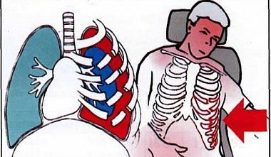
Surgical thoracoscopy. Thoracoscopy for chest trauma and injuries.
E.I. Sigal, R.G. Khamidullin. Most patients with closed and penetrating wounds to abdominal organs undergo diagnostic thoracotomy or conservative treatment. Therefore, on the one hand, unnecessary traumatic thoracotomies may be performed, while on the other, conservative tactics may delay the patient's necessary surgical intervention. Videothoracoscopy, combining minimal trauma with extensive diagnostic capabilities, allows for the selection of the optimal strategy. In many cases, the diagnostic procedure can become therapeutic. A distinction is made between emergency (performed less than 24 hours after the injury) and delayed thoracoscopy. Indications: Penetrating chest wounds below the level of the 7th rib (possibility of thoracoabdominal injury). Penetrating chest wounds projecting the heart and major vessels. — Closed chest injuries with blood loss exceeding 1 liter per day, diagnosed during thoracentesis or drainage of the pleural cavity. — Clotted hemothorax. — Pneumothorax. Contraindications: — Hemorrhagic shock. — Cardiac tamponade. — Obliteration of the pleural cavity. Preoperative examination of patients includes a physical examination (determining the presence and location of wounds, clinical signs of shock, rib fractures, etc.), chest X-ray (signs of pneumothorax, hemothorax, hemopericardium, etc.). Surgical technique. The first thoracoport for inserting the thoracoscope is inserted into cavities V-VI and, to determine the scope of the proposed intervention, 1-3 additional thoracoports are installed for inserting instruments into the pleural cavity. The insertion points and the diameter of the thoracoports used are determined depending on the location of the injury and the nature of the surgical intervention. After inserting the thoracoscope and manipulators into the pleural cavity, the pleural cavity is examined. If blood is present, the pleural cavity is drained. Diaphragmatic injury. A penetrating wound of the diaphragm revealed during examination is sutured intra- or extracorporeally. The hole in the diaphragm can be repaired through a laparotomy approach. In any case, with a thoracoabdominal injury, damage to the abdominal organs should be excluded by performing a diagnostic laparoscopy or laparotomy. Lung injury. In case of lung injury, hemostasis and pneumostasis are achieved using one of the following methods: - Application of a Roeder loop to the damaged area. - Application of a hand suture. - Stapling with a stapling apparatus. Injury to the intercostal and internal mammary arteries. Bleeding from these vessels is stopped by electrocoagulation, application of an extracorporeal suture, or clipping. Injuries to the pericardium, major vessels, bronchi, and esophagus are indications for emergency thoracotomy. Clotted hemothorax is diagnosed. Thoracoscopy is performed, and loose blood and clots are removed. The source of bleeding is determined—intercostal and internal mammary vessels, lung tissue, or rib fragments. Bleeding is stopped with electrocoagulation, clipping, or suturing. The pleural cavity is irrigated with isotonic sodium chloride and drained.


