

Use of navigation systems in endoscopic endonasal interventions
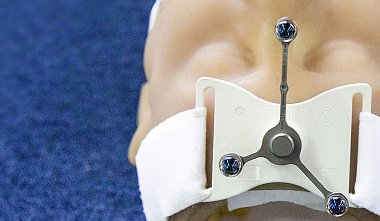
D.N. Kapitanov*, A.S. Lopatin**, A.A. Potapov* N.N. Burdenko Neurosurgery Research Institute, Russian Academy of Medical Sciences* Central Clinical Hospital, Department of Presidential Administration, Ministry of Health of the Russian Federation** Moscow Nasal endoscopy can be successfully used to remove foreign bodies from the nasal cavity and paranasal sinuses that have entered there during trauma, as well as a number of tumors that extend into the paranasal sinuses and nasal cavity. Such fragments and formations are often located at the border of the intra- and extracranial space, while direct neurosurgical intervention carries the risk of extensive damage to the structures of the skull base, including the possibility of developing basal cerebrospinal fluid leakage. Intraoperative navigation plays a significant role in such operations. Modern navigation systems allow the surgeon to navigate the complex anatomy of the surgical field during surgery and perform surgical manipulations with minimal trauma to surrounding tissues. The advent of fast portable computers and advances in imaging (MRI and CT) have enabled a technological breakthrough in stereotactic surgery. Modern frameless navigation systems have emerged, allowing surgeons to quickly and comfortably navigate the three-dimensional space of a surgical wound with an accuracy of 1-2 mm during any operation. The essence of frameless navigation technology is as follows: 1. Using an MRI or CT scanner, the surgeon receives a set of patient images 1-2 days before the operation; 2. A graphics station, which is one of the main components of the navigation system, constructs a three-dimensional image of the area to be operative. The surgeon selects reference points on the patient's head that are accessible for identification based on the resulting image (e.g., the tip of the nose, teeth, curves of the auricle, etc.); 3. Just before the operation (after induction of anesthesia), a special navigation frame (with a row of LEDs) is firmly attached to the patient's head at some distance from the surgical site. Unlike classic stereotaxis, this frame can be attached virtually anywhere and in any manner suitable for the given operation. This frame (or more precisely, a semi-arc) does not obscure the surgical field and does not restrict the surgeon's actions. 4. Next, using a special pointer with an emitter (LED), "registration" is performed, i.e., the system "links" the three-dimensional image from its memory with the actual position of the patient's head. Technically, this is accomplished using a receiver antenna, a pointer, a reference semi-arc, and a computer algorithm for working with the three-dimensional image. 5. Now, at any moment during the operation, the surgeon can control the position of their instrument with an accuracy of 1-2 mm, plan the access trajectory, and reach the selected point in the most optimal and minimally invasive manner. In the last decade, navigation systems have become widely used in intranasal procedures for tumors of the paranasal sinuses and skull base, disseminated polyps, rhinocerebrospinal fluid leakage, and other conditions. From 1999 to 2005, 16 patients (12 men and 4 women) were operated on at the N.N. Burdenko Neurosurgical Research Institute of the Russian Academy of Medical Sciences using both the endoscopic endonasal technique and the Stealth Station and Vector Vision intraoperative navigation systems. The primary patient selection criterion for intraoperative navigation included changes in the normal anatomy of the nose, paranasal sinuses, orbit, and skull base structures. The preoperative diagnosis included either tumor involvement of the aforementioned structures or a post-operative nasal condition at other medical institutions. In one case, a foreign body (bullet) located in the ethmoid labyrinth at the skull base was removed. In 10 cases, formations in the paranasal sinuses, skull base, and orbit were removed. In 4 cases, the navigation system was used during endoscopic surgeries on the paranasal sinuses in patients with multiple previous interventions in the nasal cavity, and in 1 case, a patient with nasal cerebrospinal fluid rhinorrhea was treated. A case of a 14-year-old child, admitted to the N.N. Burdenko Neurosurgery Research Institute of the Russian Academy of Medical Sciences with the diagnosis of "blind penetrating bullet wound of the orbit. Foreign metal body (bullet) in the ethmoid labyrinth on the right. Blunt trauma to the right eyeball," is presented as an example of the simultaneous use of a navigation system and endoscopic endonasal technology. The child was diagnosed with a blind penetrating bullet wound of the orbit. A foreign metal body (bullet) in the ethmoid labyrinth on the right. Blunt trauma to the right eyeball." The patient's condition was satisfactory upon admission. There were no neurological symptoms. No pathology was detected in the ENT organs, and there were no signs of nosebleeds. Computed tomography and radiography revealed a metallic foreign body in the projection of the roof of the ethmoidal labyrinth, measuring 0.5 x 0.8 x 0.8 cm, directly adjacent to the cribriform plate. Under general anesthesia, the foreign body was removed endonasally from the ethmoidal labyrinth. A combination of intraoperative navigation and endoscopic guidance allowed us to avoid a wide opening of the ethmoidal labyrinth and limit the procedure to the creation of a small channel beginning in the anterior middle meatus and leading to the base of the skull. After visualization, the bullet was grasped with forceps and removed. The accuracy of the navigation system during this procedure was 0.8 mm.


