

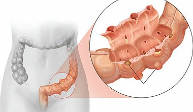
The role of laparoscopy in the treatment of acute diverticulitis
18.02.2016 15:42:00
I.V. Fedorov Kazan State Medical Academy The incidence of diverticulosis has been increasing in recent years, primarily among young people [1]. The severity of the disease ranges from mild forms to perforated diverticulitis. Over the years, the risk of perforation and other complications increases with each new relapse. In this regard, the European Association of Endoscopic Surgeons (EAES) unanimously proposed at its 1999 conference to perform elective resection of the sigmoid colon after two episodes of acute diverticulitis, or after one episode in young patients, in immunocompromised patients, or in the presence of complications such as stenosis or fistula [2]. However, recent in-depth studies of the nature of diverticular disease dictate a more gentle approach to treatment tactics. Acute complicated diverticulitis, which is the leading cause of urgent surgery, is the most common primary manifestation of the disease [3]. Recent studies show that episodes of recurrent diverticulitis have a low complication rate in patients previously treated conservatively for acute attacks of inflammation [3,4]. Accordingly, planned, prophylactic resection of the sigmoid colon after several episodes of acute inflammation is not always indicated. New diagnostic and therapeutic methods improve the results of treatment of diverticular disease. The possibility of treating abscesses with percutaneous drainage under computed tomography (CT) control shows that emergency surgery can be postponed in many cases, and planned resection can be performed according to indications [5]. The rise of laparoscopic surgery in the 1990s reduced mortality and complication rates compared to the open approach in performing planned colon resections [6]. The question of whether laparoscopy is applicable to patients with perforated diverticulitis and diffuse peritonitis remains controversial. Laparoscopic sigmoid colon resection cannot always be performed due to severe inflammatory infiltrate, fecal or purulent contamination of the abdominal cavity, severe patient condition, or comorbidities. The Hartmann procedure remains the procedure of choice for many surgeons. However, several studies show that primary anastomosis with or without a discharging stoma can be successfully performed even in such circumstances [4]. Laparoscopic lavage and drainage may be a successful alternative to the Hartmann procedure in cases of perforated purulent diverticulitis if an endosurgical approach is not contraindicated [7]. CT (with water-soluble contrast) is indicated for patients admitted with acute diverticulitis. CT is currently replacing barium enema for confirming the diagnosis in patients with suspected diverticulitis. The value of CT scanning in acute diverticulitis lies in its ability to confirm the diagnosis and classify the severity of the disease, differentiating mild localized inflammation from severe inflammation with abscess formation and/or diffuse peritonitis [5]. Before the advent of CT, contrast enema was the primary diagnostic test for colonic diverticulosis. Today, CT has largely replaced barium enema as the imaging technique of choice in patients with suspected diverticulitis. The most recent large prospective study, conducted in Switzerland, included 423 patients examined over a 10-year period [38]. The sensitivity of CT was excellent (97%). Moreover, detection of severe disease (presence of abscess or extraluminal air/contrast) was very useful prognostically and accurately predicted failure of drug therapy and the risk of secondary complications. Overall, considering the issue of cost, availability and local experience, the presented data show that if the patient is in severe condition, the diagnosis is in doubt or there is clinical deterioration, then computed tomography becomes the most appropriate initial examination method. The effectiveness of CT has been confirmed in many studies, demonstrating high sensitivity (97%) and specificity (100%) in diverticulitis [5]. Contrast enema in this situation is sensitive only in 82% of cases, and specific in 81% [10]. The patient after hospitalization should be classified according to the severity and extent of the disease, based on clinical data and the Ambrosetti classification based on CT results [2, 5]. Hinchey et al. proposed a classification of the magnitude intestinal perforation by degrees [40]. Hinchey 1 characterizes a precolytic abscess; Hinchey 2a indicates a distant (e.g., pelvic) abscess requiring percutaneous drainage; Hinchey 2b indicates a complex abscess with or without a fistula. Diffuse peritonitis is classified as Hinchey 3 (purulent) or 4 (fecal) [2, 11]. The Hinchey classification can only be used during the operation, when the stage of the destruction process (fecal or purulent peritonitis) is clear. In emergency surgery with Hinchey 3 and 4, it is necessary to approach the selection of patients for laparoscopic access extremely carefully (compliance, stability, absence of immunodeficiency and septic shock), identifying a group requiring surgery by laparotomy access (multiple comorbidities, instability, septic shock); the Hartmann operation is preferable for these patients. Elective laparoscopic surgery is a reasonable alternative to laparotomy in Hinchey stage 1 with an abscess up to 5 cm and Hinchey 2a, after percutaneous drainage of the abscess [23]. B. Franzato et al. reported on 1348 laparoscopic colectomies performed over 16 years [39]. Of these, 286 were for diverticular disease. A total of 83 (29.02%) had complicated disease, the remaining 203 (70.98%) were uncomplicated. Patients according to the Hinchey classification, group IV, were excluded, since the operation was immediately performed through a laparotomy approach or with a transition to laparotomy. All patients admitted with suspected diverticulitis underwent CT with intravenous contrast. All elective patients underwent minimally invasive surgery after a diagnostic examination. Emergency patients were treated laparoscopically, except for those who were not suitable for the procedure - hemodynamically unstable and unable to tolerate pneumoperitoneum, or having cardiopulmonary contraindications to the laparoscopic approach. The study included all patients suffering from diverticular disease, including diverticulosis, acute diverticulitis, and chronic diverticulitis. All patients underwent laparoscopic colectomy with unprotected primary anastomosis or laparoscopic lavage drainage. All colectomies were performed by the authors directly laparoscopically. Assistance was not used. A ureteral stent was not used routinely. Uncomplicated cases were defined as simple symptomatic diverticulitis resistant to medical therapy, recurrent or chronic diverticulitis, as well as non-inflammatory complications of diverticulosis. Indications for planned laparoscopic treatment were recurrences of severe diverticulitis (two or more) with radiographically proven changes. in the colon (asymmetry, wall thickening, stenosis) and/or complications of the disease after previous conservative treatment. Indications for urgent laparoscopic treatment were patients with CT and clinical data of peritonitis, who were suitable, stable and without signs of septic shock. Nine patients (3.14%) underwent urgent laparoscopic lavage and drainage for purulent peritonitis (Hinchey 3), 5 of whom were operated on 4-6 weeks later - they underwent planned resection of the sigmoid colon. The remaining four underwent this operation later. B. Franzato et al. performed 247 left-sided colectomies, 28 sigmoidectomies, 1 subtotal colectomy, 1 ileocecal resection and 9 laparoscopic lavage and drainage procedures. All patients underwent an unprotected primary anastomosis, except 4 cases of unloading ileostomy. Closed aspiration drainage was used only in particularly complex cases or when the quality of the anastomosis was in doubt. The average operative time was 160.57 min (80-330) for left colectomy and 143.79 min (80-247) for sigmoidectomy. The operative time stabilizes after completing the learning curve. The conversion rate was 1.75% (5/286), as a result of standardization of the technique and appropriate selection of patients for laparoscopy. The reason for conversion, as a rule, was the extent of the disease (1 - Hinchey 4, 2 - severe inflammation, 2 - difficulties in exposing the surgical field). The average length of the resected intestine was 28 cm (16-40). Histological examination revealed 3 carcinomas (1.04%). There was no intraoperative mortality and only one death on the 4th day after surgery from pulmonary embolism arteries. Postoperative complications occurred in 37 patients (12.97%). One bleeding from the anastomotic suture line formed with a stapler, which required transanal revision. This patient subsequently developed stenosis of the anastomosis, which required endoscopic dilation. One patient developed peritonitis on the 7th day after surgery due to covered perforation of the ileum, which required reoperation. One patient developed postoperative intra-abdominal bleeding, which was treated conservatively with red blood cell transfusion. Suppuration of the minilaparotomy wound developed in 16 (5.59%) patients. Anastomotic leakage occurred in 6 (2.09%) patients, which was treated with laparoscopic reoperation, peritoneal lavage, drainage and ileostomy. The average hospital stay was 6.74 (5-22). Long-term follow-up (on average, 48 months) did not reveal recurrence of diverticulitis, anastomotic stricture, urinary or reproductive dysfunction [39]. Laparoscopic surgery is becoming increasingly common in the planned treatment of sigmoid diverticulosis. Several studies confirm the feasibility and safety of the laparoscopic approach in this pathology [12-15]. Laparoscopic sigmectomy reduces the time of recovery, restoration of bowel function, length of hospital stay [16-18], cost [18,19], compared with open surgery. Experienced surgeons report a conversion rate of 2.8% and a hospital stay of 4 days [15]. The incidence of serious complications such as intra-abdominal abscesses, anastomotic leakage, myocardial infarction, pulmonary embolism is also lower [6]. In cases Hinchey 2b, not subject to percutaneous drainage, or Hinchey 3 of any location, or in the presence of pus in the free abdominal cavity, the laparoscopic procedure should be limited to washing and drainage without bowel resection and stoma formation [20]. In this case, there is no need to perform a wide laparotomy. Sanation laparoscopy and the administration of broad-spectrum antibiotics are sufficient [21-23]. After some time, these patients undergo planned laparoscopic bowel resection and the entire treatment becomes minimally invasive. In addition to avoiding laparotomy and stoma, this tactic allows for bowel resection in the most favorable planned situation [1, 24]. Totally minimally invasive access serves as a good alternative at stage Hinchey 3 in a carefully selected group of patients. However, there is a risk of early reoperation in cases where persistent fistulization prevents closure of the diverticular defect [24]. For this reason, it is important to have strict criteria for obtaining good results: patients in whom the location perforation is clearly visualized during laparoscopy or should be included in the Hinchey 4 group, should be excluded from the lavage drainage group [7, 21, 25-27]. These patients can have the intestine with the perforation zone brought out or undergo resection with or without a unloading stoma [26]. Some authors in this situation suggest suturing the perforation hole and/or covering it with a strand of omentum or fibrin glue [26] with good results, a low number of complications and immediate improvement in the patient's condition [22, 23]. Patients in whom a full examination of the abdominal cavity is impossible due to adhesions or obstruction, or when fecal peritonitis is detected during examination, conversion and standard resection are indicated [21,22,26] (Hartmann's operation, or resection with anastomosis with or without the creation of a unloading stoma). After peritoneal lavage and drainage, planned resection of the sigmoid colon can be performed after 3-6 months. This tactic is very promising in terms of reducing mortality and complication rates [21]. Some authors write that lavage and drainage can become the definitive method in the treatment of diverticulitis. Thus, Myers et al. [7] found a recurrence of sigmoid diverticulitis in only 4 of 92 patients, none of whom required reoperation after an average of 36 months. Such information comes from different surgical centers [28, 29], laparoscopic lavage in selected patients can become a determining method in the treatment of perforated diverticulitis [3]. However, the number of such studies is limited and is mainly based on small groups of patients, the problem requires further study. An alternative to laparoscopic lavage in Hinchey 3-4 may be laparoscopic resection in carefully selected patients. The choice between the Hartmann procedure and resection with anastomosis with with or without the imposition of a discharging stoma is decided individually in each case [30], depending on the patient's condition and the operator's skills, keeping in mind that the Hartmann procedure is preferable in patients with concomitant pathology, as well as when the quality of the inflamed tissues does not guarantee the safety of the anastomosis. There is not yet sufficient data to recommend laparoscopic resection in patients of Hinchey groups 3-4. Even in specially selected patients and in experienced hands, the effectiveness and safety of the intervention cannot be fully guaranteed [3]. Mortality and the number of complications in patients with anastomosis are similar to those in patients who have undergone the Hartmann procedure [4]. This fact confirms that in specially selected patients, surgery with anastomosis for acute diverticulitis is quite safe, even against the background of fecal peritonitis. However, the number of such observations is small [31-34], and the frequency of additional radiological interventions is high, as is the conversion rate [27]. There is not yet enough data to recommend a laparoscopic approach for routine treatment diverticular disease complicated by perforation. Therefore, prudence dictates excluding the laparoscopic approach in obviously unsuitable, unstable patients with symptoms of septic shock, or those classified as ASA classes 3-4. These patients should be referred immediately for laparotomy. Conclusion: Upon admission of a patient with acute diverticulitis, a CT scan should be performed immediately to select patients with a mild course of the disease, distinguishing them from severely ill patients (Ambrosetti classification). This first step is very helpful in better determining the indications for emergency or elective surgery, or for conservative management with or without percutaneous abscess drainage. If necessary, such abscess drainage can be performed followed by elective bowel resection and primary anastomosis - after the acute inflammation has subsided. If percutaneous drainage is unsuccessful, laparoscopic lavage and drainage of the abdominal cavity is a good alternative to urgent bowel resection in selected patients with possible subsequent planned laparoscopic bowel resection. The best way to avoid intra- and postoperative complications is to avoid surgery in the presence of acute inflammation, with the exception of patients in Hinchey groups 3-4, as well as patients resistant to conservative therapy. It should be remembered that laparoscopic planned resection is the best choice for the treatment of chronic diverticular disease [33, 35, 36]. Laparoscopic lavage and drainage are quite legitimate in the treatment of stable, suitable patients Hinchey 3 in the absence of visible and significant perforation with fresh peritonitis, with an eye to definitive planned bowel resection. If such conditions are not present, or if laparoscopic examination reveals Hinchey 4, conversion to laparotomy for bowel resection is indicated. In selected cases, such resection can be performed laparoscopically [31-34], but recommendations should be extremely cautious. For Hinchey 3 or 4, the choice between операцией Хартмана и первичной резекцией с анастомозом (с разгрузочной стомой или без неё) в каждом случае решается индивидуально, исходя из здравого смысла в зависимости от состояния пациента и мастерства хирурга. Отказ от анастомоза в пользу операции Хартмана показан нестабильным больным с множественными сопутствующими заболеваниями, страдающим иммунодефицитом, или тем, у которых качество воспалённых тканей не гарантирует безопасности анастомоза [37]. Что касается хирургической техники, показано наложение анастомоза без натяжения, что достигается полноценной мобилизацией селезеночного угла ободочной кишки. Дистальная диссекция продолжается до ректосигмоидного перехода, что предотвращает низкий рецидив дивертикулита в последующем. Лапароскопическая резекция с удалением поражённого воспалением сегмента толстой кишки (но не всех дивертикулов) может быть рекомендована [8, 9]. На основании данных литературы можно считать, что лапароскопическое лечение осложнённых форм дивертикулярной болезни даёт хорошие результаты с низкой летальностью и небольшим числом осложнений. Лапароскопия нуждается в специфической хирургической стратегии, особенно в осложнённых случаях (абсцесс, свищ). Хирургическая команда должна быть опытна как в лапароскопической, так и в колоректальной хирургии, технику операции необходимо стандартизировать, чтобы частота осложнений, время операции и число конверсий было минимальным. References 1. Stocchi L (2010) Current indications and role of surgery in the management of sigmoid diverticulitis. World JGastroenterol 16:4-17. 2. EAES (1999) EAES Consensus Statement - Diagnosis and treatment of diverticular disease. Surg Endosc 13:430-436. 3. Chapman JR, Dozois EJ, Wolff BG et al (2006) Diverticulitis: a progressive disease? Do multiple recurrences predict less favourable outcomes? Ann Surg 243:876-880. 4. Salem L, Flum DR (2004) Primary anastomosis or Hartmann's procedure for patients with diverticular peritonitis? A systematic review. Dis Colon Rectum 47:1953-1964. 5. Ambrosetti P, Jenny A, Becker C et al (2000) Acute left colonic diverticulitis compared performance of computed tomography and water-soluble contrast enema: prospective evaluation of 420 patients. Dis Colon Rectum 43:1363-1367. 6. Klarenbeek BR, Veenhof AA, Bergamaschi R et al (2009) Laparoscopic sigmoid resection for diverticulitis decreases major morbidity rates: a randomized controlled trial:short-term results of the Sigma Trial. Ann Surg 249:39-44. 7. Myers E, Kavanagh D, Hurley M et al (2008) Laparoscopic peritoneal lavage for generalized peritonitis due to perforated diverticulitis - A feasible alternative. Dis Colon Rectum 51:13. 8. Sartori CA, Balduino M, Dal Pozzo A et al (2003) Laparoscopic colonic resections for diverticular disease. Results of a single center series of 105 patients. Surgical strategy and technique. Osp Ital Chir 9:111-120. 9. Sartori CA (2007) Trattato di tecnica chirurgica laparoscopica del colon e del retto. SVP press, Padua. 10. Lawrimore T, Rhea J (2004) Computed tomography evaluation of diverticulitis. J Intensive Care Med 19:194-204. 11. Hinchey EJ, Schaal PGH, Richards GK (1978) Treatment of perforated diverticular disease of the colon. Adv Surg 12:85-109. 12. Pugliese R, Di Lernia S, Sansonna F et al (2004) Laparoscopic treatment of sigmoid diverticulitis: a retrospective review of 103 cases. Surg Endosc 18:1344-1348. 13. Schwandner O, Farke S, Fischer F et al (2004) Laparoscopic colectomy for recurrent and complicated diverticulitis: a prospective study of 396 patients. Langenbecks Arch Surg 389:97-103. 14. Garrett KA, Champagne BJ, Valerian BT et al (2008) A single training center's experience with 200 consecutive cases of diverticulitis: can all patients be approached laparoscopically? Surg Endosc 22:2503-2508. 15. Jones OM, Stevenson AR, Clark D et al (2008) Laparoscopic resection for diverticular disease: follow-up of 500 consecutive patients. Ann Surg 248:1092-1097. 16. Faynsod M, Stamos MJ, Arnell T et al (2000) A case-control study of laparoscopic versus open sigmoid colectomy for diverticulitis. Am Surg 66:841-843. 17. Dwivedi A, Chahin F, Agrawal S et al (2002) Laparoscopic colectomy vs. open colectomy for sigmoid diverticular disease. Dis Colon Rectum 45:1309-1315. 18. Lawrence DM, Pasquale MD, Wasser TE (2003) Laparoscopic versus open sigmoid colectomy for diverticulitis. Am Surg 69:499-504. 19. Senagore AJ, Duepree HJ, Delaney CP et al (2002) Cost structure of laparoscopic and open sigmoid colectomy for diverticular disease: similarities and differences. Dis Colon Rectum 45:485-490. 20. Mutch MG (2010) Complicated diverticulitis: are there indications for laparoscopic lavage and drainage? Dis Colon Rectum 53:1465-1466. 21. O'Sullivan GC, Murphy D, O'Brien MG, Ireland A (1996) Laparoscopic management of generalized peritonitis due to perforated colonic diverticula. Am J Surg 171:432-434. 22. Franklin ME Jr, Dorman JP, Jacobs M, Plasencia G (1997) Is laparoscopic surgery applicable to complicated colonic diverticular disease? Surg Endosc 11:1021-1025. 23. Da Rold AR, Guerriero S, Fiamingo P et al (2004) Laparoscopic colorrhaphy, irrigation and drainage in the treatment of complicated acute diverticulitis: initial experience. Chir Ital 56:95-98. 24. Toorenvliet BR, Swank H, Schoones JW et al (2010) Laparoscopic peritoneal lavage for perforated colonic diverticulitis: a systematic review. Colorectal Dis 12:862-867. 25. Bretagnol F, Pautrat K, Mor C et al (2008) Emergency laparoscopic management of perforated sigmoid diverticulitis: a promising alternative to more radical procedures. J Am Coll Surg 206:654-657. 26. Faranda C, Barrat C, Catherine JM, Champault GG (2000) Two stage laparoscopic management of generalized peritonitis due to perforated sigmoid diverticula: eighteen cases. Surg La-parosc Endosc Percutan Tech 10:135-141. 27. Taylor CJ, Layani L, Ghusn MA, White SI (2006) Perforated diverticulitis managed by laparoscopic lavage. ANZ J Surg 76:962-965. 28. Karoui M, Champault A, Pautrat K (2009) Laparoscopic peritoneal lavage or primary anastomosis with defunctioning stoma for Hinchey 3 complicated diverticulitis: results of a comparative study. Dis Colon Rectum 52:609-615. 29. Favuzza J, Friel J, Kelly JJ et al (2009) Benefits of laparoscopic peritoneal lavage for complicated sigmoid diverticulitis Int J Colorectal Dis 24:797-801. 30. Rafferty J, Shellito P, Hyman NH, Buie WD (2006) Practice parameters for sigmoid diverticulitis. Dis Colon Rectum 49:939-944. 31. Agaba EA, Zaidi RM, Ramzy P et al (2009) Laparoscopic Hartmann's procedure: a viable option for treatment of acutely perforated diverticulitis. Surg.Endosc 23:1483-1486. 32. Martel G, Bouchard A, Soto CM et al (2010) Laparoscopic colectomy for complex diverticular disease: a justifiable choice? Surg Endosc 24:2273-2280. 33. Scheidbach H, Schneider C, Rose J et al (2004) Laparoscopic approach to treatment of sigmoid diverticulitis: changes in the spectrum of indications and results of a prospective, mul-ticenter study on 1,545 patients. Dis Colon Rectum 47:1883-1888. 34. Zdichavsky M, Granderath FA, Blumenstock G et al (2010) Acute laparoscopic intervention for diverticular disease: a feasible approach. Langenbecks Arch Surg 395:41-48. 35. Gervaz P, Inan I, Perneger T et al (2010) A prospective, randomized, single-blind comparison of laparoscopic versus open sigmoid colectomy for diverticulitis. Ann Surg 252:3-8. 36. Pavlidis TE, Pavlidis ET, Sakantamis AK (2010) Current management of diverticular disease of the colon. Tech Coloproctol 14:79-81. 37. Tonelli F, Di Carlo V, Liscia G, Serventi A (2009) La malattia diverticolare del colon: quan-do e come trattarla. Consensus Conference 5° Congresso Nazionale della Societa Italiana dei Chirurghi Universitari (SICU). Ann Ital Chir 80:3-8. 38. Ambrosetti P, Grossholz M, Becker C, et al. (1997) Computed tomography in acute left colonic diverticulitis. Br J Surg; 84:532-4. 39. Boris Franzato, Stefano Mandala, Grazia Fusco and Carlo Sartori (2012). In: The Role of Laparoscopy in Emergency Abdominal Surgery. Springer-Verlag, Italia, . P. 77-87. 40. Hinchey EJ, Schaal PH, Richards MB. Treatment of perforated diverticular disease of the colon. (1978). Adv Surg;12:85-109.


