

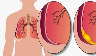
Thoracoscopy
Thoracoscopy as a diagnostic method was first used by the Swedish physician Jacobaeus in 1910 to assess the condition of the pleural cavity in tuberculosis patients. Subsequently, it was most frequently used in phthisiology to transect adhesions after the formation of therapeutic pneumothorax. The introduction of effective anti-tuberculosis drugs in the 1950s led to a decline in interest in this method. Only a small number of clinics continued to use thoracoscopy, primarily as a diagnostic tool for pleural pathology, chest trauma, and inflammatory diseases of the pleura and lungs. It was only in the early 1990s that the development of video technology revolutionized this diagnostic and treatment method. Since then, not only the surgeon but also all assistants could observe the pleural cavity, allowing for both diagnostic examinations and full-scale surgical interventions. The rapid development of video-assisted thoracoscopic surgery continues today, and only now is a real analysis of the feasibility of certain interventions being carried out, since the ability to perform an operation thoracoscopically does not necessarily guarantee a successful outcome. Equipment and instruments The following are required for video-assisted thoracoscopic surgeries: 1. Video equipment; 2. Endoscopes and video ports; 3. Stapling devices; 4. Thoracic instruments (lung clamps, dissectors, retractors); 5. Various devices for tissue coagulation (electrocoagulators, lasers, argon units). Due to the possibility of conversion to open surgery, a set of instruments for open surgery should always be ready. Video equipment. There are no significant differences in the requirements for video equipment compared to laparoscopy. The only difference may be the increased need for a second monitor, which is connected to the main monitor by a cable and placed in front of the assistant, allowing the entire surgical team to monitor the progress of the operation without leaving the surgical field. Endoscopes and thoracoports. Most surgeries are performed using a 0° straight-ahead telescope. A 30° lateral telescope can be useful for operations on the anterior and posterior mediastinum. In addition to standard 10 mm telescopes, 5 mm telescopes are now available. Newer equipment is the videothoracoscope, a device in which the video camera chip is located at the distal end of the telescope, which significantly improves image clarity. Flexible thoracoscopes, which resemble fiberscopes but have a more rigid end, allowing for a comprehensive examination of the pleural cavity, are less readily available due to their high cost. Cannulas used for thoracoscopy (thoracoports) differ from those used in laparoscopy, as thoracoscopy does not require a hermetic seal of the pleural cavity or the maintenance of a pneumothorax. Flexible thoracoports, which allow the insertion of curved instruments into the pleural cavity, are particularly attractive. Staplers are also available. The optimal solution is to use endoscopic staplers, originally developed for creating intestinal anastomoses (Endo-GIA). These devices are available in lengths from 30 to 60 mm and staple depths of 2.5, 3.5, and 4.8 mm. When activated, the stapler cuts the adducted tissue between the triple row of staples. This makes this device very useful for dissecting the lung parenchyma to ensure reliable hemostasis and pneumostasis. Unfortunately, due to their high cost, these devices are used much less frequently than desired. A more economical approach is to use standard UO-type staplers. However, their insertion into the pleural cavity requires a wider access (up to 5-6 cm in length). Instruments. Most instruments currently used for video-assisted thoracoscopy were developed for laparoscopic surgery and do not fully meet the needs of thoracic surgery. Among special instruments, curved instruments (clamps, dissectors, scissors, etc.) should be noted. Their curvature allows access to any area of ??the pleural cavity. However, their insertion is only possible through flexible plastic thoracoports or through mini-incisions. The ability to operate through mini-incisions, without the use of thoracoports, allows for the use of a wide range of traditional surgical instruments, both standard and slightly modified for videothoracoscopy, during VATS procedures. Tissue coagulation devices are also used. Traditional electrocoagulation and cutting are most commonly used. Lasers, especially lasers, are useful. Their use allows for the resection of lung nodules located on a flat surface, making staplers difficult to use. An argon coagulator is also desirable in the operating room during VATS procedures, particularly for hemostasis of diffusely bleeding surfaces of the chest wall and lung during decortication. Diagnosis and Treatment of Pleural Diseases. Thoracic surgery itself began with the treatment of pleural diseases (empyema). The first description of thoracic surgery belongs to Hippocrates, who diagnosed empyema of the pleura and cured the patient by draining the pus through an incision (nowadays this would be called thoracostomy). - Anatomy and physiology of the pleural cavity - Diagnosis of pleural pathology - Pleurisy - Empyema of the pleura - Empyema of the pleura and bronchopleural fistula - Spontaneous pneumothorax and pneumomediastinum Rare pleural infections - Pathology of the thoracic duct and chylothorax - Mesothelioma of the pleura and less common pleural tumors - Fibrothorax and decortication. The most effective method for diagnosing and treating pleural pathology in most cases is video-assisted thoracoscopy. Thoracoscopic lung biopsy. Diagnostic thoracoscopic lung biopsy is usually performed for interstitial lung disease or suspected disseminated tumor. Typical symptoms of interstitial lung disease include shortness of breath on exertion and a dry cough. Chest radiography and computed tomography (CT) findings can be quite characteristic. In disseminated tumors, shortness of breath and cough may be absent, while CT reveals more pronounced lesion nodularity, including diffuse changes combined with nodularity. Thorascopic diagnosis and treatment of mediastinal diseases. Video-assisted thoracoscopy is optimal for tumors and cysts of the posterior and medial mediastinum, especially given their predominantly benign nature. Thoracoscopic thymectomy techniques for myasthenia gravis have also been developed. It should be noted, however, that due to the significant invasiveness of thymomas, the detection of a thymoma in combination with myasthenia gravis during preoperative CT scanning requires a more cautious approach to the choice of surgical method. Surgeries for mediastinal cysts can always be performed thoracoscopically. To prevent cyst recurrence, complete removal is desirable. However, in the presence of dense adhesions between the cyst walls and surrounding structures, excision of the accessible walls and coagulation of the inner lining may be sufficient. Video-assisted thoracoscopy is essential in diagnosing the causes of enlarged intrathoracic (mediastinal) lymph nodes. Thorascopic sympathectomy and splanchnicectomy. Video-assisted thoracoscopic sympathectomy is an example of the "gold standard" of thoracoscopic surgery. While traditional sympathectomy is considered a rather traumatic intervention, endoscopic thoracic sympathectomy can be performed practically on an outpatient basis, and bilaterally. Splanchnicectomy for malignant abdominal pain syndrome caused by pancreatic cancer is also a highly effective operation due to the low trauma of the intervention.


