

Thorascopic surgery. General provisions.
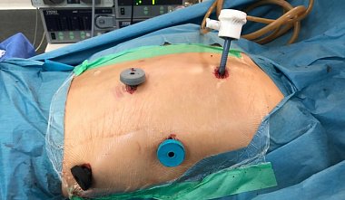
E.I. Sigal, R.G. Khamidullin The introduction of endoscopic surgery into clinical practice has also extended to thoracic operations. The previously known diagnostic thoracoscopy has developed into a new direction — operative thoracoscopy. Indications for diagnostic and operative thoracoscopy: Diagnostic thoracoscopy — Exudative pleurisy of unknown etiology. — Disseminated lung diseases. — Lung cancer staging. — Mediastinal lymphadenopathies. — Malignant tumors of the mediastinum. — Clarification of the nature of pericarditis. Operative thoracoscopy — Spontaneous pneumothorax as a complication of bullous disease. — Empyema of the pleura. — Bronchiectasis. — Peripheral benign lung tumors. — Solitary pulmonary metastases. — Peripheral lung cancer T1-2 N0. — Benign tumors of the mediastinum. — Operations on the autonomic nervous system. — Trauma and wounds of the chest. — Benign diseases of the esophagus. — Pulmonary echinococcosis. — Breast cancer (in this case, parasternal lymphadenectomy is possible). Currently, the indications for the use of thoracoscopic interventions are expanding. — Cardiac surgery (ligation of the patent ductus arteriosus, implantation of pacemaker electrodes, coronary artery bypass grafting). — Esophagectomy. — Spondylitis of various etiologies. — Pulmonary emphysema. Contraindications are usually divided into general and local. General contraindications include: — Acute myocardial infarction. — Acute cerebrovascular accident. — Uncorrectable coagulopathy. — Intolerance to one-lung ventilation. Local - obliteration of the pleural cavity. Preoperative examination — General clinical examination (complete blood count with white blood cell count, urinalysis, blood biochemistry, ECG). — Chest X-ray. — Chest tomography. — Chest CT scan. — Bronchoscopic examination. — Spirometry. Patient position on the operating table. The main requirements are the ability of the lung on the side opposite the operation to be performed to be able to function normally and ease of surgical manipulation. Most often, operations are performed with the patient lying on the healthy side. This position, although convenient for the surgeon, has its drawbacks. Compression of the healthy lung when disconnecting the lung on the side being operated on from ventilation adversely affects respiratory function. In addition, this position increases the risk of aspiration of bronchial secretions into the airways of the healthy lung, followed by the development of complications. A more gentle patient position is the semi-lateral one on a wedge-shaped bolster. In this case, the healthy lung is subjected to less compression, while the surgical field is large enough for thoracoscopy. Sometimes it is necessary to tilt the operating table to one side or the other, so the patient must be securely immobilized. In rare cases (during esophagectomy), the patient is placed prone. Anesthesia. General anesthesia with artificial ventilation is most often used for thoracoscopy, although diagnostic operations can also be performed under local anesthesia. Separate bronchial intubation with the lung on the affected side excluded from breathing provides sufficient space for the intervention. A collapsed, immobile lung creates optimal conditions for performing manipulations on both the lung itself and the organs of the chest cavity. Pneumothorax. Unlike the abdominal cavity, the chest has a bony framework, so thoracoscopy does not require the creation of an artificial space by pumping gas under pressure. A small cavity volume of approximately 200 cm3 is initially sufficient for the intervention. A sealed pleural cavity is not required for thoracoscopy, but some surgeons prefer to perform diagnostic thoracoscopy on a sealed pleural cavity with preliminary CO2 insufflation at a pressure of no more than 8 mmHg. This, however, can lead to mediastinal shift, reduced venous return, and cardiac dysfunction. There are several methods for creating a pneumothorax and inserting thoracoports: - The pleural cavity is punctured with a Veress needle in the 5th-7th intercostal space along the midaxillary line. After ensuring the correct position of the needle (air is drawn into the pleural cavity through the cannula), 200-300 cm3 of gas is insufflated. A thoracoport for the thoracoscope is then inserted into the pleural cavity at the puncture site. Puncture and gas insufflation are not performed; instead, a small skin incision approximately 1 cm long is made and the thoracoport is inserted into the pleural cavity. If adhesions or pleural obliteration are suspected, a skin incision is made, the intercostal muscles are bluntly separated, and a finger is inserted into the pleural cavity, followed by insertion of a thoracoport. A 3-4 cm mini-thoracotomy is performed in the fourth or fifth intercostal space in the projection of the anterior axillary line. Instruments (including general surgical instruments) are inserted through the created opening, and the specimen is removed through it at the end of the surgery. Most often, the first thoracoport is inserted into the fifth intercostal space along the midclavicular line. For interventions on the lower mediastinum, the third or fourth intercostal space is used, while for operations on the anterosuperior mediastinum, the sixth or eighth intercostal space is used. After examination of the pleural cavity, additional manipulation thoracoports are inserted under visual guidance. Their number and insertion sites determine the nature of the proposed surgery. For diagnostic thoracoscopy, a single thoracoport is usually sufficient. During operative thoracoscopy, two or three are inserted. Surgical Technique: Thoracoscopy is performed using a rigid thoracoscope with a viewing angle of 0° or 30°. The latter option is more convenient, as it allows for examination of the entire pleural cavity. The lung, visceral and parietal pleura, diaphragm, and mediastinal organs are examined sequentially. Attention is paid to the presence of effusion, rashes, or deposits on the pleura, and pathological formations in the mediastinum. After the operation, the lung is inflated under visual control. The pleural cavity is drained with one or two tubes inserted at the locations of the upper and lower thoracoports (Fig. 2). Postoperative Period: Patients are mobilized immediately after the end of sedation. A control chest X-ray is performed on the first postoperative day. Non-narcotic analgesics are administered as indicated. Drains are usually removed 2-3 days after surgery. The indication for their removal is the restoration of pneumostasis and the cessation of exudation (less than 100 ml per day). Complications and their prevention. Complications are divided into two groups: non-specific complications, which arise during any thoracic surgery, regardless of the method used, and specific complications during thoracic ablation. Complications of the first group include respiratory and cardiovascular failure, postoperative bleeding, wound infection, and incomplete pneumostasis. Cardiac failure and infectious complications occur less frequently during thoracoscopic procedures. This can be explained by the less invasive nature of the surgery and minor damage to the soft tissues of the chest wall. However, incomplete pneumostasis is a more common complication during thoracoscopic procedures than during open surgery, due to the more complex intracorporeal suturing technique. Bleeding during thoracoscopic ablation sometimes requires a switch to thoracotomy. Complications of the second group include injury to the diaphragm, pericardium, lung, and intercostal vessels during thoracoport insertion; mediastinal shift associated with tension pneumothorax; intercostal neuralgia; and tumor cell implantation at thoracoport insertion sites. Causes of complications and their prevention. Bleeding from large vessels of the pulmonary root during lobectomy has now become a rare complication due to improved surgical technique and the quality of medical instruments. During biopsy, selecting an avascular site and preliminary puncture of the mediastinal tumor reduces the risk of bleeding. The use of blunt-ended thoracoports and their insertion under visual control help prevent damage to the diaphragm, pericardium, lung, and intercostal vessels. Mediastinal shift is prevented by performing the surgery in a depressurized pleural cavity without CO2 insufflation. Separate bronchial intubation with the lung on the affected side excluded from respiratory function creates optimal conditions for the surgery. Intercostal neuralgia is a relatively rare complication of thoracoscopy. Its occurrence can be prevented by correct insertion of a thoracoport or the use of small-diameter flexible trocars. Implantation metastasis is prevented by using endoscopic nets and specimen removal containers. These methods eliminate tumor contact with the wound surface. Reasons for switching from endoscopic to open surgery — Reasons unrelated to the surgical technique: — Intolerance to single-lung ventilation. — Obliteration of the pleural cavity. Anatomical features (large size of the pathological lesion, fusion of interlobar fissures). Inability to visualize the pathological lesion. These situations are not considered complications of thoracoscopy. In some cases, even a thorough preoperative examination, including CT, does not provide information about a possible adhesion process and accurately determine the location of the lesion in the lung. Technique-related reasons: - Uncontrolled bleeding. - Damage to the esophagus and bronchial trunks. - Excessive duration of the surgical procedure if unsuccessful. Prolonged attempts to perform the surgery through an endoscopic approach lead to intraoperative complications. Unreasonable prolongation of the surgical procedure complicates the postoperative period and completely negates the advantages of minimally invasive techniques.


